
Please click on the tables and figures to enlarge


Exploring the environmental impact of the NHS dental service with a focus on conscious sedation and anaesthesia


B. Revert*
Speciality Doctor in Restorative Dentistry, Croydon University Hospital, 530 London Rd, Thornton Heath CR7 7YE
*Correspondence to: Dr Bethany Revert
Email: Bethany.revert@nhs.net
Revert B. Exploring the environmental impact of the NHS dental service with a focus on conscious sedation and anaesthesia. SAAD Dig. 2024: 40(II): 105-109


Abstract


Understanding the extent to which our health service contributes to the climate crisis is vital. All areas of healthcare are currently environmentally unsustainable due to being highly resource intensive. Undertaking dental treatment in both primary and secondary care contributes to the carbon footprint of our health service. This comprises the use of pharmacological intervention with both conscious sedation and general anaesthesia. This paper aims to analyse how we can continue excellent patient care while being more sustainably aware.


Introduction


Over the years, the narrative surrounding climate change has evolved and is becoming widely viewed as the biggest global health threat of the 21st century.1 The principles of beneficence and non-maleficence which underpin dental practice raise an important question: ‘How can we provide dental care without harming the environment and in turn protect our patients?’

Emerging studies have identified an increase in mental ill health and anxiety since the COVID-19 pandemic.2 The need for anxiety management within both primary and secondary care settings has never been more essential but the additional requirements of these patients will increase the carbon footprint of our services. To fully comprehend this impact, evaluation of our healthcare service delivery becomes paramount.

One of the essentials of environmental impact analysis is the quantification of the greenhouse gases. Greenhouse gases are grouped into a common unit called carbon dioxide equivalent (CO2e).3 In 2008, the UK Climate Change Act set out a legal framework to cut gas emissions to 100% below 1990 levels by 2050. This highlighted the need for collaborative effort to ensure positive and productive change to reduce the environmental footprint. High figures for CO2e and waste generation suggest that healthcare delivery is currently not environmentally, socially or financially sustainable.4 To tackle this, the NHS published a report in 2020 providing an updated account of the latest NHS carbon footprint. This report includes an ambition to achieve a ‘net zero’ health service and the initial aim is to reduce greenhouse gas emissions by 80% before 2040. Not only is this an enormous target, but the NHS was the first health service in the world to propose this.5

Most emissions from the NHS fall into three main categories: direct emissions from on-site healthcare, indirect emissions from the generation of purchased energy and indirect emissions due to producing and transporting goods. Areas of healthcare that fall outside of these main categories include staff and patient travel and the carbon footprint of prescribed medication.5

Accurately quantifying the carbon footprint of dental services is challenging. However, it is suggested that 3% of the overall carbon footprint of the NHS in England is due to dental services, which equates to a total carbon footprint of around 670,000 people.6,7

Within the 2021 Lancet report on health and climate change, it was stated that current global commitments are insufficient. They estimated that with no changes we will see a 2.4°C global temperature increase by the end of the century. If the global temperature were to rise above 2°C this would lead to worldwide consequences, including death due to intense heat, alterations in food availability and increased infectious disease transmission.8


Dentistry as a whole


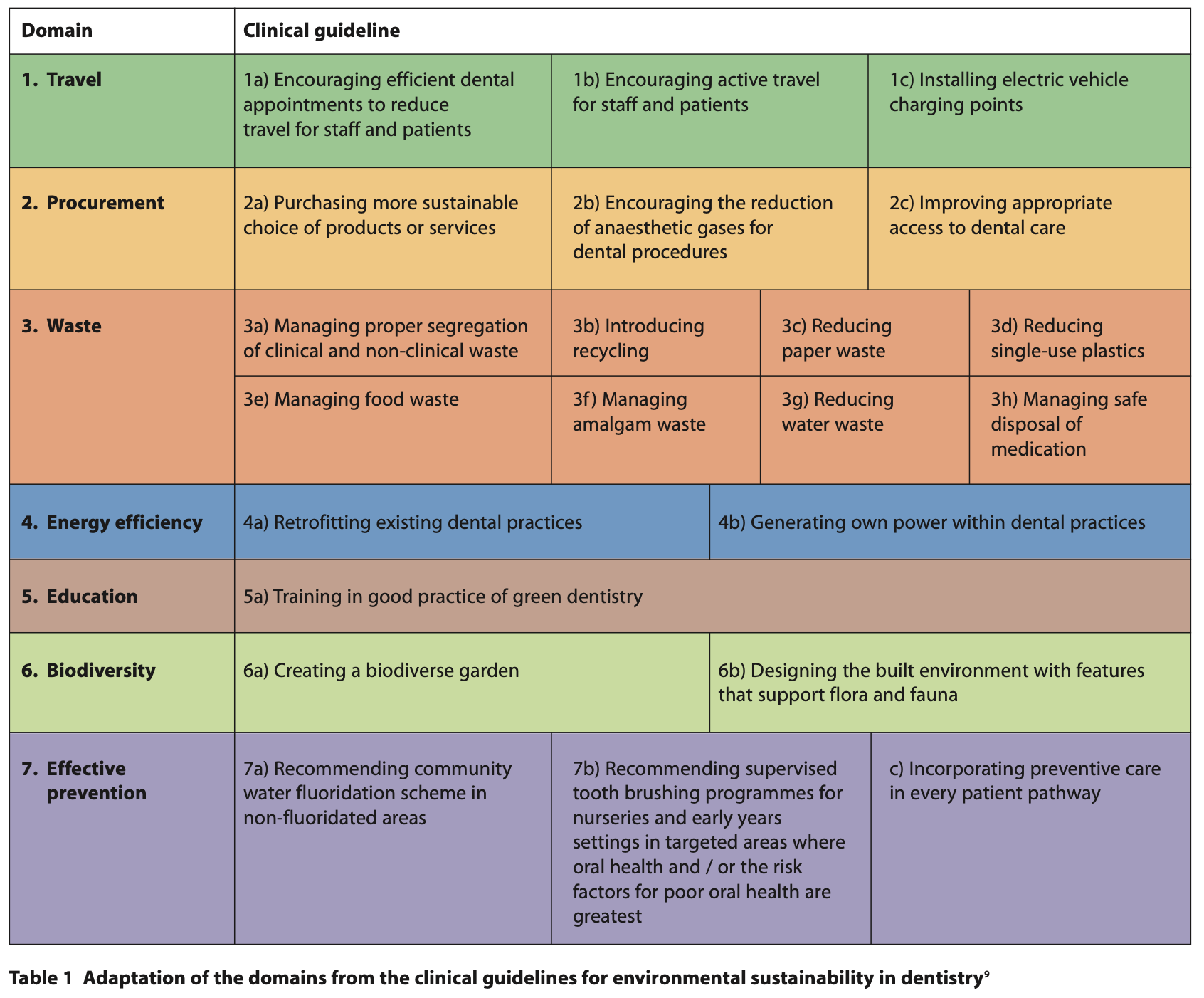
With the joint pressure of the NHS Net Zero target and the Climate Change Act, dentistry within the UK has had to further investigate its role in reducing our carbon footprint. In January 2023, clinical guidelines for environmental sustainability in dentistry were published. These were the first dentistry-focused guidelines relating to improving the sustainability of our profession. The overall aim of these guidelines is to raise awareness, provide needed direction to the dental cohort and to reduce carbon emissions.9 These guidelines incorporate the 2030 United Nations (UN) sustainable development goals, which emphasise the need to ‘take urgent action to combat climate change’.10

Within the clinical guidelines are seven major domains, which can be seen below in Table 1. Each of these breaks down the multiple facets that lead to the overall carbon footprint within dentistry in England. These guidelines are relevant for individual practitioners, governments and public health professionals. Domain two directly mentions the reduction of anaesthetic gases for dental procedures.9

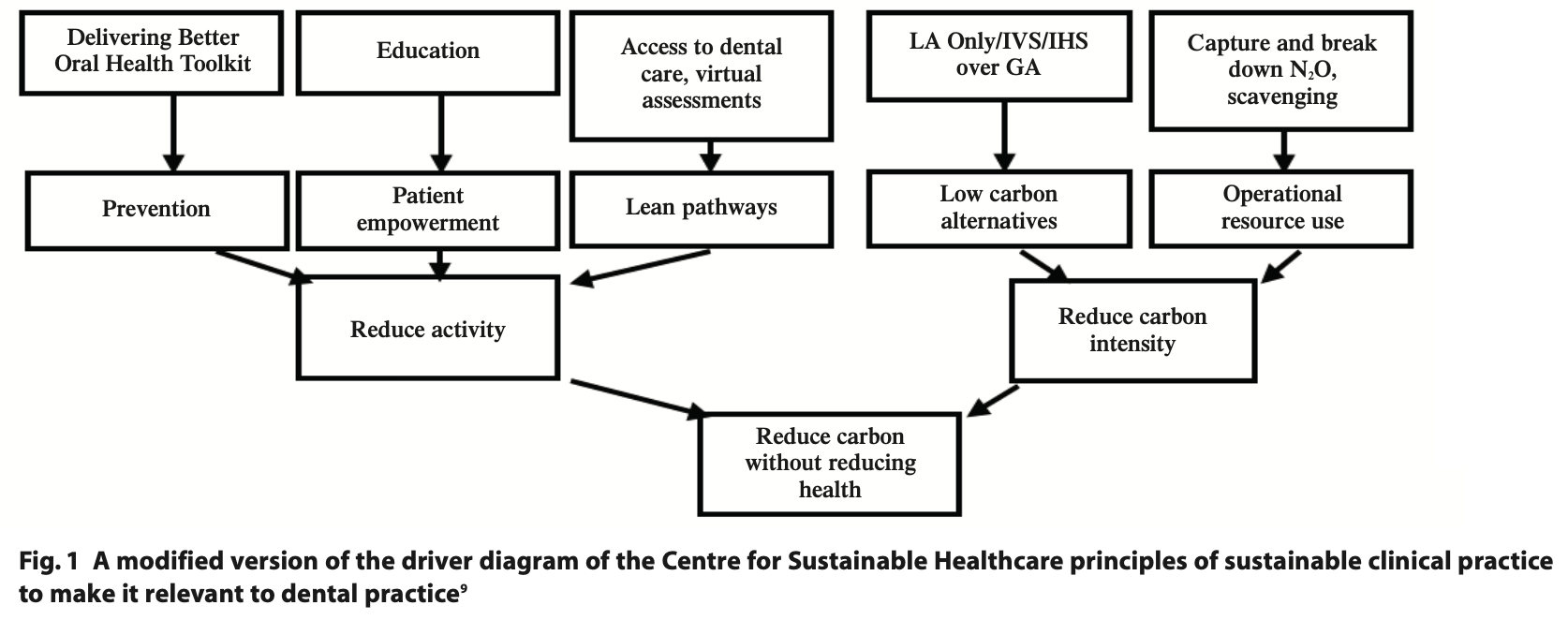
Another useful tool is the Campaign for Greener Healthcare’s four principles. These include underpinning sustainable practice, including disease prevention and health promotion, patient education and empowerment, lean service delivery and preferential use of treatment with a lower environmental impact.11 These areas of improvement can be seen illustrated in the driver diagram in Figure 1.

To be able to reduce carbon intensity, we must investigate which procedures have the largest carbon footprint. Examinations, scale and polishes, radiographs and fluoride varnish applications have the lowest carbon footprint due to the minimal materials used. However, due to the extensive number of examinations required, it still equates to a large proportion of our carbon footprint. Procedures with higher carbon footprints come from more extensive procedures, which either require increased material use or multiple appointments. In clinical situations where conscious sedation or general anaesthesia (GA) is required, an increased carbon footprint occurs due to the addition of more staff and the use of N2O or other gases.7

The most used dental materials are amalgam and composite. Excluding the presence of mercury within amalgam, the comparative carbon footprint of these two materials is unknown. In addition, the environmental breakdown of these products is not understood. Motivation for these analyses to take place is required, allowing the profession and public to make an informed material choice. However, it is known that dental mercury equates to 3 - 4% of terrestrial mercury. In total 75 metric tonnes of amalgam are used per year in the EU alone.3 The Minamata Convention has highlighted the need to reduce mercury use across the globe, which has directly impacted usage in dentistry. Conversely, in clinical situations where moisture control is difficult to achieve or when applying the most affordable option, amalgam is still a material of choice.12

Although looking at dental procedures and materials is important, we know that the highest proportion of emissions due to dentistry in the UK came from travel, procurement, energy and N2O.7 Therefore, further analysis of indirect carbon usage in dentistry needs to be completed. Approximately 3.5% of all road travel in England is due to healthcare-related excursions.5 Despite it being an indirect cause, encouraging staff and patients to switch to lower carbon footprint transport options is essential. Alongside this, we can evaluate the patient pathway by highlighting access to dental services. NHS England is required to ensure it commissions services in areas that are easily accessible for patients to travel to and from, as well as being available to all.7 Timely diagnosis and treatment, can minimise the need for further invasive treatment. There is a delicate undertaking with this, as more practices also add to increased output and a higher carbon footprint.

In the same way that access is important, we must also explore how an integrated care model could improve our carbon footprint.13 Certain areas of the UK don’t have facilities for dental treatment under sedation, therefore in some circumstances, GA may be the only option for these patients. GA requires more staff and increased use of ozone-depleting gases. Therefore, if it was possible to design and deliver more efficient, effective patient pathways we could reduce the need for unnecessary high carbon footprint procedures. 

To tackle both travel and the use of resources, one suggestion to make dentistry more sustainable is to complete as much treatment in one session as possible. However, when pharmacological intervention is required due to dental anxiety or medical history, there is a limiting factor of drug working time. Whilst this may be more achievable under GA, this is not necessarily the most practical or safe option due to theatre availability and the risks of anaesthesia. Furthermore, GA itself carries a heavy carbon burden through travel, staffing, enormous waste generation and energy consumption.14

Before using pharmacological interventions, all steps should be taken to try the least invasive option. This can include acclimatisation of dental treatment. If this is not possible due to severe anxiety, an appropriate pre-assessment should be completed. This includes an anxiety assessment tool, such as the Modified Dental Anxiety Scale.15 Patients are required to reach a certain threshold to qualify for sedation techniques under our current health service. This is a useful tool in ensuring we are only undertaking appropriate treatment measures. To reduce the environmental impact of sedation even further, reducing the prevalence of dental anxiety is required. Ideally, the use of psychotherapeutic interventions should be first-line management before pharmacological agents. However, in patients with severe anxiety, a combination of both may be required. The use of these additional psychological interventions could lead to a patient being able to undergo dental treatment without conscious sedation thereby improving patient outcomes, whilst reducing the environmental footprint as a side effect.16


Inhalation sedation with N2O


In current dental practice inhalation sedation (IHS) is undertaken inboth primary and secondary care settings. Indications for its use include dental anxiety, needle-phobia, traumatic or prolonged dental procedures, medical conditions aggravated by stress and patients who require sedation and who have no escort.17

In dentistry, we regularly use nitrous oxide (N2O) for conscious sedation which is a potent greenhouse gas. The global warming potential (GWP) of N2O is 298 times higher than CO2. Despite it having a shorter life cycle than CO2, the damage it causes within the same timeframe is much increased.18 The N2O released during the sedation of an estimated 63,749 patients, produced almost 1% of the total carbon footprint of NHS dental services in England.8

In dental practice, the exhaled gas is removed using a scavenging system. This delivers the exhaled vapours out of the clinical environment, which aims to reduce the occupational exposure of staff. It is common practice to also ensure adequate fitting of the nasal hood to ensure no excess N2O is released into the local environment.7 However, N2O does not undergo any form of metabolic change within the human body, and therefore any exhaled gas will pollute the planet.3 Suitable alternatives to releasing this exhaled N2O are being explored. Technological advances are being trialled, mainly within theatre settings in the UK, though European counterparts have already been using N2O denaturing devices for several years.

Despite N2O’s high GWP the use of IHS is seen as a less invasive option compared to intravenous sedation (IVS) or GA. Therefore, there is a delicate balance between minimising the use of IHS while still offering the least invasive treatment option.7


General anaesthesia using ozone-depleting gases


It has been estimated that an average of 163 litres of N2O is used during one procedure of GA per patient episode. This does not consider the carbon footprint of the travel and procurement process.3 The use of inhalational anaesthetic agents equates to around 5% of the total carbon footprint of acute NHS trusts. It is therefore unsurprising that when N2O is used, it equates to the biggest component of the carbon footprint of GA.19

Steps can be taken to reduce the amount of N2O used during GA. These include ensuring well-fitting masks, flushing of the system correctly, turning off valves as soon as no longer required and ensuring there are no leaks within the system.3

The use of catalytic splitting could cut over one-third of NHS anaesthetic emissions.5 This process requires an external unit to heat the N2O to 400°C, producing both oxygen and nitrogen as by-products. This technology has been successfully deployed in Sweden.20 Currently this is more readily used for GA due to the ease of installing one large unit for multiple theatres. However, we are starting to see the use of smaller units in dental settings for single rooms. For example, Medclair has produced a mobile destruction unit which reduces over 99% of N2O to its harmless byproducts.21 This is currently being piloted in England to investigate the ease of use for dental services. Promising results have been seen with this technology, for example a 52% decrease in N2O emissions from birthing units in Stockholm.20

As healthcare professionals, we already aim to try and avoid the need for GA due to additional medical risks. The knowledge of the increased environmental impact of GA allows us to further warrant the use of less invasive means. Due to the recent COVID-19 pandemic, waiting lists for GA suites are high and ever-growing,22 therefore further funding is required to allow the implementation of such technologies to offset the high use of theatres.


Sedation using midazolam and alternatives


Where IHS is contraindicated or anxiety management is unsuccessful, alternative pharmacological interventions can be applied. One of the most common options is the use of midazolam. The route of delivery for this drug can be either intranasal, oral or intravenous. Generally intranasal and oral routes are used pre-operatively before completing cannulation for intravenous midazolam use.23

Compared to non-sedation related practices, we can be certain the carbon footprint of the additional drug and environmental impact for disposal will be greater. However, comparing IVS directly to inhalation practice is more complex. It has been suggested that total intravenous anaesthesia could have a lower environmental impact, it also reduces the risk of occupational exposure and pollution to the atmosphere. Despite this, correct disposal of all IV agents should be followed to reduce environmental contamination.24 This evidence could be applied to IVS, however, within dentistry this is seen as the more invasive option over IHS. This is due to the prolonged recovery period, the requirement for an escort and the additional monitoring required. Therefore, despite the possible reduced carbon footprint, we are unable to ethically suggest the use of intravenous midazolam over IHS. 

In certain situations, for example severe dental anxiety or low co-operation, midazolam can be administered alongside other agents or alternative agents used entirely. These include fentanyl, propofol, ketamine and sevoflurane gas.23 Sevoflurane is less environmentally damaging compared to N2O and it is comparable for sedation, but further research and adaptation to dentistry is required before routine use.25 

Like midazolam there is limited evidence on the environmental impact of fentanyl, propofol and ketamine use for dentistry. These drugs are used as advanced techniques and are likely occurring in small numbers.23 Further research is required to fully understand the breakdown of these products in the ecosystem.


Clinician education


A recent study showed that only 9% of students believed they had formal teaching on sustainability despite 97.3% of students believing the profession should be more actively engaged.26

For many years, the General Medical Council has implemented specific learning outcomes for undergraduates, focusing on sustainable healthcare.27 This was an early sign that interest in the link between our medical practices and the environment was increasing. In November 2023, the General Dental Council launched the new framework for dental professional education, which will be implemented in undergraduate programmes from 2025. This framework includes a sub-domain called social accountability, requiring formal teaching on sustainable oral health and the environmental impacts of common treatment methods.28 This change allows us to engage the future generation of healthcare professionals to enable them to aim for more sustainable practice.

For our postgraduate colleagues, an introductory course on sustainability is available free for all on Health Education England’s e-Learning for Health platform.29,30 Training courses such as this require promotion by local trusts to ensure the workforce is aware of their existence.


Patient education


A downstream, preventative approach to oral health care will have the biggest impact on reducing the need for dental treatment.31 Preventative items such as fluoride toothpaste, fluoride varnish and fissure sealants can reduce the likelihood of large areas of dental caries and therefore restorative treatment.31 Education for care staff or carers can add another layer to the preventative approach. Improving education and knowledge can reduce an individual's likelihood of poor oral health. We must ensure we are following the Delivering better oral health toolkit to assess the risk status of our patients and giving them tailored advice and education.31


COVID-19


During the COVID-19 pandemic, phone triage became the only option for patients who were experiencing dental pain and swelling, and many patients were left without access to treatment.32 Despite national restrictions being lifted there has been a continuation of this practice with some using an initial video assessment before treatment.33 This is a useful tool to mitigate the need for travel for initial discussions or where a clinical exam is not required. This is useful for patients with access or transport difficulties. The long-term implementation of virtual assessments and telephone consultations could help reduce patient travel.


Conclusion


The long-term sustainability of our health service relies on the collaboration and motivation of all specialities. Successfully treating patients with conscious sedation or GA is a clinical necessity and so further research is required to allow us to aim for a more environmentally sustainable practice. Indirect causes of carbon usage such as travel, procurement and wastage require further alteration and motivation from the workforce at all levels. The use of N2O is concerning, although clinically necessary for anxiety management, and advances in catalytic cracking are likely to have the biggest impact on reducing the damage caused by N2O. Downstream prevention of both dental disease and dental anxiety is vital to help reduce the need for treatment, this practice can only be successful with patient and clinician education. In conclusion, we can shape a more sustainable and safe future by collaboratively working together.


Declaration of Interest


 There are no conflicts of interest.


Acknowledgements


I would like to acknowledge Dr Zahra Shehabi, for inspiring me to become involved in this area of dentistry, as well as her continued support in my career.


References


1. Costello A, Abbas M, Allen A, et al. Managing the health effects of climate change. Lancet 2009; 373: 1693-1733.

2. Santomauro D F, Herrera A M M, Shadid J et al. Global prevalence and burden of depressive and anxiety disorders in 204 countries and territories in 2020 due to the COVID-19 pandemic. Lancet 2021; 398: 10312: 1700–1712.

3. Martin N, Sheppard M, Gorasia G P, Arora P, Cooper M, Mulligan S. Awareness and barriers to sustainability in dentistry: A scoping review. J Dent 2021; 112: 103735.

4. Duane B, Stancliffe R, Miller F A, Sherman J, Pasdeki-Clewer E. Sustainability in dentistry: a multifaceted approach needed. J Dent Res 2020; 99: 998-1003.

5. Greener NHS. NHS England and NHS Improvement. Delivering a Net Zero NHS. 2020. Online information available at: https://www.england.nhs.uk/greenernhs/publication/delivering-a-net-zero-national-health-service/ (accessed March 2024).

6. Public Health England. Carbon modelling within dentistry: towards a sustainable future. Crown: London. 2018. Online information available at: https://assets.publishing.service.gov.uk/government/uploads/system/uploads/attachment_data/file/724777/Carbon_modelling_within_dentistry.pdf (accessed March 2024).

7. Duane B, Lee M B, White S, Stancliffe R, Steinbach I. An estimated carbon footprint of NHS primary dental care within England. How can dentistry be more environmentally stable? Br Den J 2017; 223: 589-593.

8. Romanello M., McGushin A, Napoli D et al. The 2021 report of the Lancet Countdown on health and climate change: code red for a healthy future. Lancet 2021; 398: 1619-1662.

9. Duane B, Fennell-Wells A. 2023. Clinical guidelines for environmental sustainability in dentistry.

10. United Nations DoEaSA, The 17 United Nations Sustainable Development Goals. 2015. Available at: https://sdgs.un.org/goals (accessed March 2024).

11. Mortimer F. The sustainable physician. Clin Med (London) 2010; 10: 110-1

12. Fisher J, Varenne B, Narvaez D, Vickers C. The Minamata Convention and the phase down of dental amalgam. Bulletin of the World Health Organization. 2018; 96: 436–438.

13. Willcocks S, Willcocks E. The shift to integrated care in the NHS: implications of the new care models for dentistry. Br Dent J 2019; 226: 319–322.

14. Shelton C L, McBain S C, Mortimer F, White S M. A new role for anaesthetists in environmentally-sustainable healthcare. Anaesthesia 2019; 74: 1091–1094.

15. Humphris G M, Dyer T A, Robinson P G. The modified dental anxiety scale: UK general public population norms in 2008 with further psychometrics and effects of age. BMC Oral Health. 2009; 9:20.

16. Appukuttan, D. Strategies to manage patients with dental anxiety and dental phobia: literature review. Clin Cosmet Investig Dent 2016; 8: 35–50.

17. Coulthard P. The indicator of sedation need (IOSN). Dent Update 2013; 40: 466–471.

18. Erisman J W, Galloway J, Seitzinger S, Bleeker A, Butterback-Bahl K. Reactive nitrogen in the environment and its effect on climate change. Curr Opin Environ Sustain 2011; 3; 81-290.

19. Shelton C L, McBain S C, Mortimer F, White S M. A new role for anaesthetists in environmentally sustainable healthcare. Anaesthesia 2019; 74(9): 1091–1094.

20. Ek M, Tjus K. Destruction of Medical N20 in Sweden. In Greenhouse Gases – Capturing, Utilization and Reduction. pp 187-198. London: IntechOpen, 2012.

21. Medclair. MDU - Mobile Destruction Unit Medclair. 2013. Online information available at: https://www.medclair.com/en/mdu (accessed March 2024).

22. NHS. Delivery plan for tackling the COVID-19 backlog of elective care. 2022. Online information available at: https://www.england.nhs.uk/coronavirus/wp-content/uploads/sites/52/2022/02/C1466-delivery-plan-for-tackling-the-covid-19-backlog-of-elective-care.pdf (accessed March 2024).

23. The dental faculties of the Royal Colleges of Surgeons and the Royal College of Anaesthetists. Standards for Conscious Sedation in the Provision of Dental Care (V1.1), Report of the Intercollegiate Advisory Committee for Sedation in Dentistry. 2020. Online information available at: https://www.saad.org.uk/IACSD%202020.pdf (accessed March 2024).

24. Varughese S, Ahmed R. Environmental and Occupational Considerations of Anesthesia: A Narrative Review and Update. Anesth Analg 2021; 133: 826-835.

25. Allen M, Thompson S. An equivalence study comparing nitrous oxide and oxygen with low-dose sevoflurane and oxygen as inhalation sedation agents in dentistry for adults. Br Dent J. 2014; 217: E18–8.

26. Durnall, O., Martin, N., Mulligan, S., Dixon J. Environmental sustainability: the attitudes and experiences of UK students in the oral health care profession. Br Dent J 2024.

27. Duane B, Harford S, Ramasubbu D et al. Environmentally sustainable dentistry: a brief introduction to sustainable concepts within the dental practice. Br Dent J 2019; 226: 292–295.

28. General Dental Council. The Safe Practitioner: A framework of behaviours and outcomes for dental professional education, dentist.2023. Available at: https://www.gdc-uk.org/docs/default-source/safe-practitoner/spf-dentist.pdf?sfvrsn=c198211d_5 (accessed March 2024).

29. Fennell-Wells, Amarantha. The current status of sustainability in dentistry: a perspective. Br Dent J 2023; 234: 245. Doi: 10.1038/s41415-023-5566-6.

31. Public Health England. Delivering better oral health: an evidence-based toolkit for prevention. 2014. Online information available at: https://www.gov.uk/government/publications/delivering-better-oral-health-an-evidence-based-toolkit-for-prevention (accessed March 2022).

32. Hurley S, Neligan M. Letters, updates, and additional guidance for dental teams. 2020. Online information available at: https://www.england.nhs.uk/coronavirus/publication/preparedness-letters-for-dental-care/ (accessed March 2022).

33. Parker K, Chia M. Patient and clinician satisfaction with video consultations in dentistry - part one: patient satisfaction. Br Dent J 2021. Online information available at: https://www.nature.com/articles/s41415-021-3007-y#citeas (accessed March 2024).


Impact of anxiety and midazolam on physiological control of arterial blood pressure (BP) and heart rate (HR)


S. Rehman* BDS, MFDS(RCS Ed) MSc (Pub Health) DipConSed
Dental Core Trainee in OMFS, Gloucester Royal Hospital, NHS Foundation Trust, Great Western Road Gloucester England GL1 3LZ
*Correspondence to: Dr Sooda Rehman
Email: drsoodsrehman@hotmail.com
Rehman S. Impact of anxiety and midazolam on physiological control of arterial blood pressure (BP) and heart rate (HR) SAAD Dig. 2024: 40(II): 110-112


Abstract


Knowledge of human physiology and the properties of the sedative agents used in the provision of dental treatment with conscious sedation is vital. The most commonly used sedative agent for intravenous (IV) sedation is midazolam which belongs to the benzodiazepine group of drugs. Both anxiety and midazolam affect the heart rate. However, long-term anxiety could result in hypertension. Soon after its induction midazolam affects the heart rate by changing the autonomic nervous system control of heart function as well as baroreflexes. It is worth noting that heart rate during sedation with midazolam will not reach the base level, whereas blood pressure drop could be uncertain.


Introduction


An appropriate medical history, social history and physical assessment is key to ensure an appropriate case selection for conscious sedation. Therefore, knowledge of the pharmacodynamics and pharmacokinetics of the drugs that are used in sedation is essential along with their interactions with other drugs which the patient may take routinely for any existing medical conditions. According to the Adult Dental Health Survey 2009, anxiety is prevalent in over a one third of the United Kingdom’s population.1 Conscious sedation plays a pivotal role in the provision of dental treatment for patients who suffer from dental anxiety and / or dental phobia. The most commonly used sedative agents in dentistry are midazolam and nitrous oxide. Midazolam belongs to the benzodiazepine group of drugs. Like all other sedative agents midazolam depresses consciousness as well as cardiovascular function. In this paper the impact of anxiety and midazolam on physiological control of arterial blood pressure and heart rate will be discussed.


Heart rate


The heart is an organ which pumps oxygenated blood to the different parts of the body and deoxygenated blood back to the lungs. To achieve this function the heart has to contract continuously in a co-ordinated fashion.2

The number of times a heart beats in a minute is defined as the heart rate (HR).3 Normally, a heart beats around 60 to 80 times a minute.3 Major factors which can modify the HR are as follows.3


Autonomic tone


The autonomic nervous system (ANS), which comprises the parasympathetic nervous system (PNS) and the sympathetic nervous system (SNS), provides the innervation to the heart. The autonomic tone is generated through the balance between the inputs from SNS and PSN. The parasympathetic input to sino-atrial node (SAN) is dominant at rest.3


Response to pain and anxiety


The body’s response to fear, anxiety or stress triggers SNS that innervates the atrioventricular node (AVN) and SAN and releases noradrenaline, resulting in increased HR and force of contraction.

Baroreceptor mechanism, chemoreceptors and circulating hormones are discussed later in the section entitled ‘Physiological Control of Arterial blood pressure and HR’.


Cardiac output


The total volume of blood in an average adult is 5 to 6 litres.3 The volume of blood pumped by the heart in a minute is called cardiac output (CO) which is the product of the HR and stroke volume (SV).4 SV is defined as the amount of blood pumped by the left ventricle in a single contraction.5 A resting CO in an average adult is 5.5l/min.3


Peripheral resistance


To pump the blood through the circulatory system, resistance to blood flow must be overcome. The resistance generated through the systemic vascular system is known as total peripheral resistance (PR).6

The SNS controls the radius of the blood vessel, and any changes in the radius can cause a significant change in the blood pressure (BP) by altering the PR. Similarly, factors which affect the blood’s viscosity, eg temperature, haematocrit value and plasma protein concentration, also affect the BP by increasing or decreasing the PR.


Arterial blood pressure


‘Arterial blood pressure (ABP) is determined by the volume ejected by the heart into the arteries, the elastance of the walls of the arteries, and the rate at which the blood flows out of the arteries.’ 7 ABP can also be defined as a product of cardiac output and peripheral resistance.


Physiological control of arterial blood pressure and heart rate


The ABP is regulated by the following major mechanisms:

Baroreceptors

The carotid sinus and aortic arch have specialised stretch receptors called baroreceptors. These receptors detect changes in the BP and send impulses to the vasomotor centre (VMC) to regulate the BP by increasing or decreasing the HR and CO.5

Chemoreceptor reflex

As a result of tissue ischemia, peripheral chemoreceptors are stimulated which stimulates VMC and results in an increase in HR, BP and depth of respiration.5

Central nervous system (CNS) ischaemic response

CNS response is initiated when BP falls below 50mmHg and results in stimulation of the VMC which stimulates SNS to increase HR and BP to maintain normal function.5

Renal regulation of BP

The kidneys regulate arterial blood pressure through direct mechanism (ie pressure diuresis) and the pressure natriuresis and the indirect mechanism (ie hormonal control). Major hormones are as follows:5

  • Renin angiotensin system (RAS): provides the long-term control of ABP. Through this system, the kidneys compensate for the decrease in ABP by the release of a endogenous vasoconstrictor (ie angiotensin II) that raises the ABP.
  • Aldosterone: is a steroid hormone released from the adrenal cortex by angiotensin II (ATII) which increases the sodium resorption and increases the excretion of potassium in a distal convoluted tubule of the kidney resulting in an increase of BP. ATII also increases PR by vasoconstriction of the arterioles to increase BP.
  • Antidiuretic hormone (ADH): ATII also causes the release of ADH (vasopressin) from the posterior pituitary which increases the blood volume by reabsorption of water from the kidneys which increases the BP.
  • Epinephrine and norepinephrine: these hormones are released by the adrenal medulla under stress, pain and anxiety etc as a part of the ‘fight or flight’ mechanism. They raise the BP by increasing the HR and contractility of the heart muscles while causing vasoconstriction of the arteries and veins.

Thyroxine

Thyroxin is a hormone produced by the thyroid gland which increases the HR and CO by acting on beta 1 receptors and potentiates the effect of epinephrine. Other hormones like atrial-natriuretic peptide and brain natriuretic peptide also have an impact in the regulation of HR and BP.2


Effect of an anxiety / phobia on blood pressure and heart rate


Some patients are not able to accept dental treatment due to their high level of anxiety or phobia. Anxiety and fear stimulate the SNS, resulting in an increase of the circulating catecholamines and an increase in the HR and BP.

Anxiety is considered to have a short and long-term effect on the BP. In the short term it can increase the BP and ‘white coat syndrome’ is a good example of this.8 The long-term effect of anxiety can result in decreased vascular variability resulting in an increased PR which can eventually cause hypertension.8

Generalised anxiety is often associated with an unhealthy lifestyle which, in turn, could cause an increased risk of hypertension. Higher risk of death is noted due to ischaemic heart disease among patients who have phobic anxiety disorder.9


Effect of midazolam on blood pressure and heart rate


Midazolam is short acting benzodiazepine which has been used in dentistry since the 1980s to provide sedation. Administering a sedative dose of benzodiazepine will result in approximately a 5 to 10% reduction in BP. Midazolam is known to affect BP and HR by changing the ANS control of the heart function as well as baroreflexes.10

Midazolam has a depressant effect on the sympathetic response that produces a small decrease of the ABP immediately after induction by decreasing PR, myocardium contractility and venous return. However, this is promptly compensated by the baroreceptor reflex and the HR is then increased so as the myocardium contractility to maintain the CO.

This is particularly relevant as the HR will not drop to a true resting rate during sedation, a less experienced seditionist may confuse this with under-sedation. It is uncertain to what extend the fall in BP is a result of the relief of anxiety, or the action of the drug itself.

This hypotensive effect is of clinical significance amongst elderly patients or patients with compromised cardiovascular disease. Midazolam used in high doses can cause deep sedation and the direct inhibition of the cardiovascular system (CVS).11 However, in combination with other drugs in low dose it can cause CVS stability.12


Discussion


The person with systolic and diastolic blood pressure below 140/90mmHg is classed as normotensive, whereas sustained increase of systolic blood pressure measurement of 140mmHg with, or without, an increase of diastolic blood pressure above 90mmHg is defined as having clinical hypertension.13 According to the Office for National Statistics’ 2023 report based on the Health Survey for England for 2015 – 2019, 32% of the United Kingdom’s population living in private households suffered from hypertension, with 29% having undiagnosed hypertension.14 

There was reported to be a higher incidence of undiagnosed hypertension in the population aged 16 to 34 compared to the population aged over 75.14 The increased prevalence of hypertension has been associated with increased age and high BMI along with other factors like ethnicity and region etc.14 These statistics highlight the importance of pre-assessment for conscious sedation provision in detecting any undiagnosed hypertension. This could lead to better outcomes for the patient in general as it can lead to a timely management of hypertension and its causes.15 The clinician needs to adopt a holistic approach throughout the patient’s journey from pre-assessment to recovery following a procedure under conscious sedation as multiple factors like ‘white coat syndrome’, the patient’s regular medication, anxiety, inadvertent introduction of adrenaline based local anaesthetic into a vein etc could potentiate the impact of midazolam on the cardiovascular system.


Conclusion


A sound knowledge of human physiology is imperative for the safe practice of conscious sedation as it allows the operator to understand the impact of sedative agents on blood pressure and heart rate. Recording of all the basic vital signs during pre- assessment is fundamental for the patient’s safety as it helps in appropriate case selection. Conscious sedation in dentistry is a safe alternative to general anaesthetic provided it is practised by a trained seditionist. Anxiety management during the provision of dental treatment can help to reduce the risks to cardiovascular function.


Acknowledgements


 Work was carried out as part of PG diploma in conscious sedation in King’s College London.


Conflicts of Interest


 There are no conflicts of interest.


References


1. Adult Dental Health Survey: England, Wales, Northern Ireland, 2009. Theme 8: Access barriers to care. Available at: https://digital.nhs.uk/data-and-information/publications/statistical/adult-dental-health-survey/adult-dental-health-survey-2009-summary-report-and-thematic-series. (Accessed on 7.4.24).

2. Gordan R, Gwathmey J K, Xie LH. Autonomic and endocrine control of cardiovascular function. World J Cardiol 2015; 7: 204-214. Available at: DOI: 10.4330/wjc.v7.i4.204.

3. Craig D, Boyle C. Practical Conscious sedation. 2nd ed. London: Quintessence Publishing, 2017.

4. Vincent J L. Understanding cardiac output. Crit care 2008; 12: 174 Available at: doi:10.1186/cc6975 (Accessed on 07.04.24).

5. Waghmare L S, Srivastava K T. Conceptualizing physiology of arterial blood pressure regulation through the logic model. Adv Physiol Educ 2016 40: 477-479. Available at: doi:10.1152/advan.00074.2016.

6. Siddique A. Effects of Vasodilation and Arterial Resistance on Cardiac Output. J Clinic Experiment Cardiol 2011; 2: 11. Available at: DOI: 10.4172/2155-9880.1000170.

7. Magder S. The meaning of blood pressure. Crit Care 2018: 22: 257 Available at: https://ccforum.biomedcentral.com/articles/10.1186/s13054-018-2171-1 (Accessed on 07/04/24).

8. Pan Y, Cai W, Cheng Q, Dong W, An T, Yan J. Association between anxiety and hypertension: a systematic review and meta-analysis of epidemiological studies. Neuropsychiatr Dis Treat 2015; 11: 1121-30. Available at: doi: 10.2147/NDT.S77710.eCollection 2015.

9. Gorman J M, Sloan R P. Heart rate variability in depressive and anxiety disorders. Am Heart J. 2000; 140: 77-83.

10. Win N N, Fukayama H, Kohase H, Umino M. The different effects of intravenous propofol and midazolam sedation on hemodynamic and heart rate variability. Anesth Analg 2005; 101 :97-102,

11. Watanabe Y, Higuchi H, Ishii-Maruhama M et al. Effect of a low dose of midazolam on high blood pressure in dental patients: a randomised, double-blind, placebo-controlled, two-centre study. Br J Oral Maxillofac Surg. 2016; 54: 443-8. Available at: doi: 5 10.1016/j.bjoms.2016.02.006.

12. Choi Y F, Wong T W, Lau C C. Midazolam is more likely to cause hypotension than etomidate in emergency department rapid sequence intubation. Emerg Med J. 2004; 21: 700-2.

13. Leader R, Thayer T, Maher B, Bell C. Hypertension - an update for the dental (sedation) team. Dental Update 2019; 46: 508-13 Available at: https://www.dental-update.co.uk/content/medicine-in-dentistry/hypertension-an-update-for-the-dental-sedation-team/. (Accessed on 07.04.24).

14. Office of National Statistics. Risk factors of undiagnosed high blood pressure in England 2015 to 2019. 2023. Available at: https://www.ons.gov.uk/peoplepopulationandcommunity/healthandsocialcare/healthandwellbeing/articles/riskfactorsforundiagnosedhighbloodpressureinengland/2015to2019. (Accessed on 07.04.24).

15. National Institute for Health and Care Excellence. Hypertension in adults: diagnosis and management. 2023. Available at: https://www.nice.org.uk/guidance/ng136/chapter/Recommendations#diagnosing-hypertension (Accessed on 07.04.24).


Please click on the tables and figures to enlarge


Is there a differential sedative effect in ADHD patients?


N. Kafil* BDS
Foundation general dentist, Royal Free London NHS Foundation Trust, Pond Street, London, NW3 2QG
*Correspondence to: Dr Nicka Kafil
Email: drkafiln@gmail.com
Kafil N. Is there a differential sedative effect in ADHD patients? SAAD Dig. 2024: 40(II): 113-116


Abstract


Attention deficit hyperactivity disorder (ADHD) is a common but relatively undiagnosed disability which may require conscious sedation to allow dental treatment to take place. Sedation is indicated due to issues with behavioural management, however, the types of sedative agents used affect the GABAergic pathways in the brain. In studies, patients with ADHD have shown a reduced concentration of gamma-aminobutyric acid (GABA). This discussion theorises whether there is a differential impact of sedation on ADHD patients. There is a lack of literature to provide sufficient evidence to allow a conclusion highlighting a research gap in the matter, with some research contradicting others. Further studies are required to provide quantitative data to form valid conclusions. This in turn will provide recommendations to practitioners so there are evidence-based improvements in sedation for patients with ADHD.


 Methodology


The methodology for the systematic literature review on dental sedation and ADHD.
The inclusion criteria consisted of:

  • studies published in peer-reviewed journals
  • research conducted on dental sedation, midazolam, nitrous oxide and ADHD
  • studies in the English language.

Comprehensive research across PubMed databases with relevant keywords: ‘ADHD’, ‘midazolam’, ‘nitrous oxide’ and ‘conscious sedation’. The relevant publications are free full text from 1970 to 2022 with studies on humans including clinical trials, meta-analysis and systematic review. Additional reference chaining by reviewing citations of key articles.


Introduction


The American Psychiatric Association Diagnostic and Statistical Manual, Fifth Edition (DSM-5-TR) defines attention deficit hyperactivity disorder as a mental disability. It presents as a persistent pattern of inattention, hyperactivity and / or impulsivity that impacts development and executive functioning.1 These characteristics can be obstacles for dental treatment to be completed, as there is often difficulty with the inactivity required from the patient to allow treatment to be undertaken successfully. Sedation could be attempted as an adjunct to behaviour management to facilitate treatment for patients with ADHD. Midazolam is a common agent used for intravenous sedation, midazolam acts alongside gamma-aminobutyric acid (GABA) to produce the sedative effect.2 However, patients with ADHD are reported to have a reduced GABA concentration. There is limited literature about the relationship between reduced GABA in ADHD patients and the sedative effect of midazolam.3 This paper aims to discuss the current evidence and theories to determine whether sedation has a differential impact on patients with ADHD.


Sedation indications for ADHD patients


A meta-analysis estimated around 5% of children and adolescents have a formal ADHD diagnosis worldwide.1 There is a disproportionate diagnosis rate of males compared to females in childhood. This figure is an estimate either due to a lack of international acknowledgement of the validity of the DSM-5-TR or disregard of diagnosis due to stigma towards the condition. Additionally, there is ambiguity in the prevalence of ADHD in adults as the DSM-5-TR is tailored for adolescent characteristic symptoms.4 Thus, the estimated percentage is not representative of the true number of patients with ADHD due to potentially a high proportion of the population being undiagnosed.

ADHD presents as a spectrum of symptoms, impacting individuals in varying intensities which may lead to behavioural disruptions and the inability to co-operate during dental treatment. There are three subtypes of ADHD: inattentive, hyperactive, or impulsive.4 Each individual with ADHD does not fall into a specific subtype butrather on a spectrum of traits. This may cause difficulty for the dental practitioner to use behaviour management techniques to prevent distractibility and restlessness to enable treatment to be carried out safely. In these instances, sedation may be considered a viable adjunct. Other indications for conscious sedation of ADHD patients are co-morbidity of anxiety and intellectual disabilities.5 

Patients with ADHD are classified as high caries risk and often require numerous dental visits. This is supported by a study in New Zealand which reported that ADHD patients were 12 times more likely to have decayed, missing and filled teeth (DMFT) than the control group.6

Blomqvist M, Ahadi S, Fernell E et al.,7 suggested that the frequency of tooth brushing for children with ADHD is 34% lower than in the control group, thus the increased number of dental visits is to allow shorter recall intervals to promote better oral health behaviours.7 Another contributing factor is the psychostimulant medications used alongside antidepressants which are typically prescribed to treat ADHD patients. These also have a common side effect of xerostomia, which contributes to the high caries risk categorisation.8

A study in South Korea investigated the yearly trend of ADHD patients undergoing dental treatment under sedation using the Korean National Health Insurance data. Figure 1 presents the annual number of ADHD patients undergoing dental treatment. It increased from 2007 to 2018 with the largest increase in sedation adjuncts being 14% in 2016. However, the number of sedation cases for ADHD patients decreased after 2016.9 Nevertheless, despite a decrease in sedation used for ADHD patients, there is still an increased need for dental treatment. This indicates that a continuation of prevention is imperative; this will aid in reducing the amount of treatment and continue to decrease the number of sedation procedures.


Dentists have a professional and ethical responsibility to provide optimal health care for individuals with special care needs. However, in some instances, the behaviour management strategies used by dentists are insufficient for patients with ADHD. Sedation could be a useful adjunct to dental treatment for ADHD patients as it reduces the risk of complications arising during treatment due to the patient’s inability to co-operate. It also helps the patient have a less anxiety-provoking experience which may improve their perception of dental treatment, which may in turn improve their oral health in the long term. Overall, the use of dental sedation may be in the patient’s best interests.


Mechanism of action


The two most common types of conscious sedation noted in this discussion are inhalation sedation (IHS) with nitrous oxide and oxygen and intravenous sedation (IVS) with midazolam. There are other variations in administration methods and sedative agents used.

Nitrous oxide is a gas that produces effects of anxiolysis, sedation and muscular relaxation, however, it is a poor anaesthetic.10 It is characterised by rapid onset and fast recovery due to a minimum alveolar concentration (MAC) of 1.04 atmospheres absolute.11 It is administered with a mixture of oxygen to ensure the patient is respiring sufficient oxygen levels. Also, levels of nitrous oxide should not routinely exceed 50% as negative side effects are more likely to occur.10

The guidelines from the Intercollegiate Advisory Committee for Sedation in Dentistry (IACSD 2015) and the Scottish Dental Clinical Effectiveness Program (SDCEP 2017) favour nitrous oxide as a type of conscious sedation.12-13 However, it requires good co-operation with behaviour management for patients without ADHD.

Therefore, this may not be the best option for patients with ADHD as it does not overcome the issue of the complexity of the behaviour management required.

The anxiolytic impact of nitrous oxide is thought to involve the activation of GABAA receptors, although it is uncertain whether nitrous oxide acts directly or indirectly upon the binding site.14 A paper by Fujinage and Maze15 discussed the impact of nitrous oxide on opioid receptors through the release of opioid peptides. This causes the activation of nociceptive processing in descending pathways located in the spinal cord. The noradrenergic descending pathway is inhibited due to the inhibition of the GABA pathway from opioid peptide release.14

There are no current studies to conclude whether this impacts an ADHD patient’s altered GABA pathway. Inhalation sedation may not be the most optimal adjunct for ADHD patients as it still requires behaviour management. Additionally, another factor is the possible GABA-dependent activity, with unknown impacts on ADHD patients. Though it may be the preferential sedation type in guidelines, nitrous oxide may not be as suitable for ADHD patients.

Midazolam is a 1,4-imidazobenzodiazepine derivative with properties of a short duration of action, rapid onset and water solubility.16 Midazolam can be administered through multiple routes: oral, intramuscular, intranasal and intravenous. There is a rapid resorption rate due to its unique structure, with a pH-dependent ring opening which causes increased lipid solubility at a physiological pH. Midazolam is bound to plasma proteins in serum albumin with clearance of midazolam through liver metabolism by P450 cytochrome.17

Midazolam has effects of sedation, anxiolysis, temporary amnesia, muscle relaxation and anti-convulsion.2 However, there are side effects including, but not limited to, respiratory depression, excessive sleepiness, dizziness, confusion, nausea and vomiting.18

The physiological effects of midazolam are due to the action of midazolam on the receptors of the neurotransmitter GABA.2 GABAA receptors are ligand-gated ion channels that control GABA transmission, which acts as an inhibitory neurotransmitter to achieve inhibition of the central nervous system.19 Midazolam increases GABA binding to GABAA receptors, increasing the effects of GABA, thus increasing the frequency of chloride channel opening. This leads to hyperpolarisation of the membrane which decreases the action potential in the post-synaptic neurone. This causes neuronal inhibition and thus the inhibition of the central nervous system.2

GABAA receptors consist of five subunits, the gamma (γ) subunit is imperative for benzodiazepine's impact on the GABAA receptor. Benzodiazepine and GABA bind to a chloride ion channel separately. However, without the γ subunit, the benzodiazepine is unable to alter membrane potential through GABAA receptor activity.20


The association of GABA with ADHD


ADHD is a developmental disability that is associated with a deficient dopamine and noradrenergic neurotransmitter system.21 A study by the American Academy of Neurology used transcranial magnetic stimulation to present a correlation between decreased short intercortical inhibition with ADHD symptoms of reduced motor control and impulsivity. Short intercortical inhibition is modulated by GABAA agonists, which is imperative for processing sensory information and utilising appropriate behavioural responses.22 Another study using magnetic resonance spectroscopy provided evidence that also supports the hypothesis that the difficulty in behaviour inhibition is linked to reduced GABA concentration.3 

Opposing literature from the New York Academy of Sciences argues that the gamma-aminobutyric acid receptor subunit theta (GABRQ) gene encoding the GABAA receptor subunit is associated with ADHD. With a variance in personal impact due to polymorphism in X-linked GABRA3 and GABRB3 genes.23 ADHD has a comorbidity with autism spectrum disorder (ASD). A study of ADHD and ASD presented that the GABRQ gene is associated with both disorders. The transgenic model noted mutations in the GABAA receptor which may be associated with reduced GABA and inhibition response. However, the GABRQ gene did not prove a significant association, suggesting a hypothesis of genetic variants across genes that produce a combined effect. Thus, additional research is necessary for clarification.24

The literature can be categorised into two main theories about the relationship between GABA and ADHD. Either there is a reduced GABA concentration or a GABAA receptor mutation in patients with ADHD. Unfortunately, none of the studies identifies whether there is reduced GABA concentration with a normal number of GABA receptors or vice versa. Additionally, the specific distribution and location of reduced GABA or GABAA receptors are not determined.3 This may be very important when considering intravenous sedation with midazolam for patients with ADHD.


Impact of sedation on ADHD patients


The impact of sedation on ADHD patients has not been studied and / or widely reported. Therefore, in this discussion the evidence in studies reporting on either theory of altered GABA released or GABAA receptor mutation is utilised to theorise the impacts.

The inhibition of neurones through GABA binding occurs in phasic and tonic forms. Phasic inhibition is controlled by synaptic receptors, resulting in the fleeting desensitisation of GABAergic conduction. Tonic inhibition occurs via extra-synaptic receptors that lead to a persistent type of GABA conductance.25 ADHD presents altered concentrations of GABA release, which causes both the phasic and tonic inhibition forms to be reduced in the amygdala; the co-ordinating centre of emotional behaviour in the brain.22

Mutation of GABAA receptor subunits leads to an ineffective response to GABA.22 Literature from Molecular Pharmacology at the University of Singapore suggests that in neurological diseases the decreased number of GABAA receptors has similar effects to the mutated GABAA receptors.27

A study on cerebral blood flow changes due to midazolam shows a dose-related reduction in regional cerebral blood flow in the cingulate gyrus, prefrontal cortex and thalamus. This causes impaired cognitive function in these areas.28 The anterior cingulate gyrus in the brain involves emotional processing and behavioural control. Additionally, it has extensive connections to the amygdala.29

From the evidence about reduced GABA concentration in the studies mentioned above it is possible to theorise that for ADHD patients there is a reduced impact from midazolam during sedation, either from decreased GABA secretion or GABAA receptor mutation. This may lead to less inhibition in the amygdala and cingulate gyrus during sedation, exhibiting reduced dampening on behaviour and emotional responses targeted in sedation. This means that whilst using midazolam, the ADHD patient is more difficult to calm and relieve their behavioural symptoms, which may interfere with dental treatment. This is also possible due to the connection between ‘emergence agitation’ and ADHD patients noted in a study by researchers at the University of Michigan.29 Emergence agitation is when patients are irrationally excited, agitated, restless and combative during early phase sedation recovery. The causative factor of emergence agitation is theorised to be due to the altered GABAA receptor activity.29 The Sachdev and Kruk model describes this as being due to decreased inhibition signals to the thalamocortical neurones, thus an increase in ADHD characteristics and less behavioural control.30

On the contrary, a study published in the Korean Journal of Anesthesiology31 reported a similar sedative effect on children with, and without, ADHD in the emergency department. The mean sedation scores recorded for each patient presented no requirements for modifications of midazolam sedation used for children with ADHD. The data presented no significant difference in sedation depth, however, there was an incidental finding of an increase in sedation duration for ADHD patients. This study had a limitation of non-standardised dosage therefore, variance could have possibly introduced differences between the cases.32 This case does not provide any input on the theory discussed previously as there are no records of whether there were any differences in the extent of ADHD symptoms presented during sedation and whether behavioural control was like patients without ADHD.

The definition and practice of conscious sedation vary between countries due to differences in health system regulations, sedation techniques and the drugs used. For instance, the maximum recommended concentration of nitrous oxide differs across European countries, ranging from 50% to 70%.33 These variations mean that success rates observed in one country cannot be directly applied to another, such as the UK. This discrepancy was highlighted in an international prospective cohort study conducted across Japan, Korea and Taiwan, emphasising that sedation practices and regulations differ globally.34 There is a limitation in the comparison of studies due to the differences in sedation practices across countries. This variation leads to challenges in assessing the impact of sedation uniformly, as different drugs and techniques may be used. This is especially important because there is very little research on sedation in patients with ADHD. The limited data available on this topic further complicates the ability to draw reliable comparisons and develop standardised guidelines for sedation practices in the ADHD patient group.


Sedation considerations for the management of ADHD patients


Patients with ADHD require both behavioural and pharmacological management to regulate cognitive abilities and behaviours. Sedation is a consideration when non-pharmacological management is not sufficient for dental treatment to occur. The use of stimulant medication is a common treatment option for ADHD, with methylphenidate (Ritalin) being the most used.6 A case report by Ririe D, Kirsten R, Navil S et al.,32 presented the difficulties of conscious sedation with oral midazolam in patients with ADHD who are medicated with Ritalin. It is a stimulant medication; therefore, it was inferred that although it decreases the behaviour impulsivity of the patient, it is potentially a reason for the difficulty in obtaining the desired sedation level. This led to increased doses and additive time required to reach the level of desired conscious sedation.32 Whilst planning for pharmacological management, the dental sedationist should consider drug interactions, as the stimulant medication could potentially counteract the sedative agents. 

Another consideration is the difference in sedative drug metabolism in ADHD patients. Ritalin impacts the liver through the inhibition of microsomal enzymes which in turn impacts metabolism and elimination.8 Midazolam is metabolised in the liver via microsomal oxidase.35 It is possible to suspect that findings of prolonged sedation, in the study in the Korean Journal of Anesthesiology,31 are due to Ritalin impacting the midazolam metabolism in the patient. The dental sedationist might consult the patient’s psychiatrist before treatment. This is because there is little literature, therefore guidelines, about the interaction between ADHD stimulant medications and sedative agents.

As previously discussed, the risk of dental caries and xerostomia is higher in ADHD patients. Therefore, it is paramount for the dental practitioner to provide a tailored prevention protocol. In turn, this will reduce the number of ADHD patients who require treatment and thus sedation.


Conclusion


There are multiple case studies and theories about the correlation between ADHD and sedation. Unfortunately, there is a lack of systematic investigations with sufficient sample sizes and set variables to prove a definitive answer.

Many of the current studies available on ADHD patients are based on children, there is an overall lack of literature on the dental management of adult ADHD patients. Most patients seen by a general dental practitioner are adults, therefore further observations and different sample groups are needed to provide guidance towards sedation treatment.

Multiple quantitative studies need to be completed for adults and children with ADHD separately, each analysing the effects of dosage of sedation types compared to a control group. This should allow a meta-analysis to be conducted to provide evidence for the theories discussed. Further studies will allow specific guidelines to be formed to help the dental sedationist and provide optimum dental care for patients with ADHD.


References


1. American Psychiatric Association. 2022. What is ADHD? Online information available at: https://www.psychiatry.org/patients-families/adhd/what-is-adhd.

2. Wang J, Sun P, Liang P. Neuropsychopharmacological effects of midazolam on the human brain. Brain Informatics. 2020; Nov 10: 7-15.

3. Edden R A, Crocetti D, Zhu H et al. Reduced GABA concentration in attention-deficit/hyperactivity disorder. Arch Gen Psychiatry. 2012;69: 750-3.

4. Polanczyk G V, Willcutt E G, Salum G A et al. ADHD prevalence estimates across three decades: an updated systematic review and meta-regression analysis. Int J Epidemiol. 2014;43: 434-42.

5. Wilens T E, Spencer T J. Understanding attention-deficit/hyperactivity disorder from childhood to adulthood. Postgrad Med. 2010;122: 97-109.

6. Broadbent J M, Ayers K M S, Thomson W M. Is attention-deficit hyperactivity disorder a risk factor for dental caries? A case-control study. Caries Res. 2004;38: 29-33.

7. Blomqvist M, Ahadi S, Fernell E et al. Dental caries in adolescents with attention deficit hyperactivity disorder: a population-based follow-up study. Eur J Oral Sci. 2011;119: 381-5.

8. Sinha S, Praveen P, Rani S P et al. Pedodontic Considerations in a Child with Attention Deficit Hyperactivity Disorder: Literature Review and a Case Report. Int J Clin Pediatr Dent. 2018;11: 254-259.

9. Chi S I, Kim H, Seo K S. Analysis of application of dental sedation in attention deficit hyperactivity disorder (ADHD) patients using the Korean National Health Insurance data. J Dent Anesth Pain Med. 2021;21: 99-111.

10. Hallonsten A L, Jensen B, Raadal J et al. EAPD Guidelines on Sedation in Paediatric Dentistry. 2003.

11. Röpcke H. Effects of nitrous oxide on mac. Best Practice & Research Clinical Anaesthesiology. 2001; 15: 409–16.

12. Intercollegiate Advisory Committee for Sedation (IACS). Standards for Conscious Sedation in the Provisions of Dental Care: Dental clinical guidance. 2015. Online information available at: https://www.saad.org.uk/IACSD%202020.pdf (accessed September 2023).

13. Scottish Dental Clinical Effectiveness Programme (SDCEP). Conscious sedation in Dentistry: Dental clinical guidance. 2022. Online information available at: https://www.sdcep.org.uk/media/iota3oqm/sdcep-conscious-sedation-guidance-unchanged-2022.pdf (accessed September 2023).

14. Emmanouil D E, Quock R M. Advances in understanding the actions of nitrous oxide. Anesth Prog. 2007;54: 9-18.

15. Fujinaga M, Maze M. Neurobiology of nitrous oxide-induced antinociceptive effects. Mol Neurobiol. 2002;25: 167-89.

16. Emmanouil D E, Quock R M. Advances in understanding the actions of nitrous oxide. Anesth Prog. 2007;54: 9-18.

17. Vasakova J, Duskova J, Lunackova J et al. Midazolam and its effect on vital signs and behaviour in children under conscious sedation in dentistry. Physiol Res. 2020;69: 305-314.

18. Ashton H. Toxicity and adverse consequences of benzodiazepine use. Psychiatric Annals. 1995;25: 158–65.

19. Wang J, Sun P, Liang P. Neuropsychopharmacological effects of midazolam on the human brain. Brain Inform. 2020;7:15.

20. Eom W, Lee J M, Park J et al. The effects of midazolam and sevoflurane on the GABA(A) receptors with alternatively spliced variants of the γ2 subunit. Korean J Anesthesiol. 2011;60: 109-18.

21. Kim Y S, Yoon B E. Altered GABAergic Signalling in Brain Disease at Various Stages of Life. Exp Neurobiol. 2017;26: 122-131.

22. Gilbert D L, Isaacs K M, Augusta M et al. Motor cortex inhibition: a marker of ADHD behaviour and motor development in children. Neurology. 2011;76: 615-21.

23. Rothman D L, Petroff O A, Behar KL et al. Localised 1H NMR measurements of gamma-aminobutyric acid in human brain in vivo. Proc Natl Acad Sci U S A. 1993;90: 5662-6.

24. Comings D E. Clinical and molecular genetics of ADHD and Tourette syndrome. Two related polygenic disorders. Ann N Y Acad Sci. 2001;931: 50-83.

25. Naaijen J, Bralten J, Faraone S et al. Glutamatergic and GABAergic gene sets in attention-deficit/hyperactivity disorder: association to overlapping traits in ADHD and autism. Transl Psychiatry. 2017;7:999.

26. Lee V, Maguire J. The impact of tonic GABAA receptor-mediated inhibition on neuronal excitability varies across brain region and cell type. Front Neural Circuits. 2014;8:3.

27. Yuan H, Low C-M, Moody O A et al. Ionotropic GABA and Glutamate Receptor Mutations and Human Neurologic Diseases. Mol Pharmacol. 2015;88: 203-17.

28. Powers W J. Cerebral blood flow measurement with positron emission tomography. Cerebral Blood Flow. 2003: 217–25.

29. Rolls E T. The cingulate cortex and limbic systems for emotion, action, and memory. Brain Struct Funct. 2019;224: 3001-3018.

28. Reddy S K, Deutsch N. Behavioral and Emotional Disorders in Children and Their Anesthetic Implications. Children (Basel). 2020;7: 253.

29. Lindenmayer J P. The pathophysiology of agitation. J Clin Psychiatry. 2000;61: 5-10.

30. Catharine A W, Dawn E, Trevor R. Behavioural models of impulsivity in relation to ADHD: Translation between clinical and preclinical studies. Clin Psychol Rev. 2006; 26: 379–395.

31. Shin Y H, Kim M H, Lee J J et al. The effect of midazolam dose and age on the paradoxical midazolam reaction in Korean paediatric patients. Korean J Anesthesiol. 2013;65: 9-13.

32. Ririe D, Kirsten R, Navil S et al. Unexpected interaction of methylphenidate (Ritalin) with anaesthetic agents. Paediatr Anaesth. 1997;7: 69-72.

33. Ashley P, Anand P, Andersson K. Best clinical practice guidance for conscious sedation of children undergoing dental treatment: an EAPD policy document. European Archives of Paediatric Dentistry. 2021; 22: 989–1002.

34. Yang C H, Chen P J, Mori M et al. Cross-cultural comparison of continuous deep sedation for advanced cancer patients in East Asian countries: prospective cohort study. Japanese Journal of Clinical Oncology. 2023; 1–9.

35. Brichard G, Johnstone M. The effect of methylphenidate (Ritalin) on post- halothane muscular spasticity. Br J Anaesth. 1970;42: 718-22.

Please click on the tables and figures to enlarge


Cannulation conundrums: what help is available?


A. Gupta*1, BDS, PG Cert, MFDS RCSPG
T. Nagpal2, BDS, PG Cert, PG Dip, MFDS RCSEd
1Oral Surgery Registrar, Southend Hospital, Prittlewell Chase, Southend-On-Sea, SS0 0RY
2Oral Surgery Registrar, Luton and Dunstable Hospital, Luton, LU4 0DZ
*Correspondence to: Ashana Gupta
Email: Ashana.Gupta2@nhs.net
Gupta A, Nagpal T. Cannulation conundrums: what help is available?. SAAD Dig. 2024: 40(II): 117-123


Abstract


A fundamental requirement of intravenous sedation is access. Successful cannulation is vital for delivery of the sedative, reversal agents and for management of medical emergencies. Experienced clinicians, however, will be able to recall occasions where they have struggled to gain intravenous access.

Pre-operatively, the consultation visit can be utilised to prepare the patient for sedation cannulation. Patients can be advised on hydration to improve vein visibility at the time of cannulation. The clinician can also visualise blood vessels that would be suitable for cannulation at the initial assessment. Application of the Adult Difficult Intravenous Access Scale to grade cannulation difficulty is useful for record keeping as well as preparing the clinician for cannulation difficulties and organising appropriate adjuncts. At the time of cannulation, tapping of the vein, placement of warm compresses, milking of veins proximally to distally and application of a tourniquet can increase venous prominence. Adjuncts that can be used include the sphygmomanometer cuff at or below diastolic pressure, transillumination and topical venodilation with nitroglycerine ointment. The availability of portable handheld ultrasound devices has also enabled ultrasound guided cannulation within a dental setting.

This paper provides an algorithm to guide the clinician when managing difficult cannulation.


Introduction


Intravenous (IV) cannula placement is required to administer sedative medication as well as reversal agents. This is often the first clinical stage of delivering conscious sedation, with the exception being those that must be given a form of premedication or alternative sedation technique to aid the acceptance of IV cannulation. It can be argued that being prepared with the full adjunctive armamentarium for successful IV access is crucial as the gold standard for delivery of the sedation reversal agents is through intravenous administration.

Cannulation can be considered a challenging skill with a cross-sectional survey conducted in 25 countries stating a potential failure rate between 31% and 67%.1 Repeated attempts at cannulation can pose a burden to both the patient and healthcare system. Risks associated with repeated cannulation include pain, infection, nerve damage, bleeding, haematoma, air embolism and occlusion.2,3

The average duration for ‘time taken to cannulate’ varies between 32 seconds and two minutes in successful cases of venous access. When access is difficult, reports indicate the time taken can increase to be between 66 seconds and five minutes.4

The Welsh peripheral intravenous cannulation best practice guidelines suggest a maximum of two failed cannulation attempts. Clinical judgement should be exercised and reassessment with senior support, or cannulation adjuncts may be needed.5 This paper aims to inform clinicians of their options to aid adult cannulation and ultimately ensure they are able to gain venous access as efficiently as possible, overcoming arguably the most crucial step to be able to deliver successful conscious sedation.


Pre-operative patient assessment


Venous access should be assessed during the patient’s consultation visit when treatment planning sedation. Consideration of the patient’s medical history is essential as this may complicate cannulation. Relative contraindications for insertion of a peripheral cannula at a particular site may include phlebitis, sclerosed veins, burns, history of trauma, obesity, ipsilateral mastectomy and lymph node dissection.2 Awareness of these factors will enable the sedationist to prepare and choose an appropriate venous access site. Swift cannulation and administration of sedation drugs is an important step in managing the anxious patient.

As part of the consent process, patients should be made aware of the need for cannula insertion, potential insertion sites, the associated risks and potential complications.6,7

The National Institute for Health and Care Excellence (NICE) recommends that clinicians make a clinical decision regarding the ideal site and choice of vein before cannulating, to reduce the risk of cannula failure and complications.8 Sites to be avoided include any veins in proximity to arteries, superficial or sclerosed veins. If the patient is postmastectomy in breast cancer treatment, the side associated with breast surgery is best avoided due to the risk of lymphoedema developing post node removal.5

The cephalic or basilic vein on the forearm (Figure 1A), cephalic vein of the wrist and the dorsal metacarpal veins on the back of the hand (Figure 1B) are considered best practice for cannulation.7


Predicting difficult venous access


Cannulation can fail for several reasons. These can be categorised to systemic patient factors, local patient factors or clinician factors as shown in Table 1.

Systemic factors

Patients assessed to be American Society of Anaesthesiologists (ASA) I and ASA II are appropriate for management in a primary dental care setting for conscious sedation, those who are considered ASA III should be referred to secondary or tertiary settings and are likely to have more difficult venous access due to a combination of the above factors.

To mitigate some of the preventable factors, appropriate pre-operative instructions should be given to patients attending for sedation to maximise the chances of successful cannulation. Fasting prior to dental conscious sedation is an often-discussed issue. As loss of airway reflexes should never occur in conscious sedation in dentistry, fasting is unnecessary.10,11 Eating and drinking appropriately allows patients to minimise the risk of dehydration prior to attending and increases the visibility of their veins. Secondary or tertiary care settings may have fasting instructions which may contrast with this due to the use of polypharmacy and broad hospital policies also covering our anaesthetic and gastroenterology colleagues and their procedures under conscious sedation.

Local factors

The Adult Difficult Intravenous Access Scale (A-DIVA) is a recently created predictive scale to identify patients with difficult intravenous access. This additive A-DIVA scale predicts the likelihood of a difficult intravenous access in adult patients prospectively, based on clinical observations and five variables (Table 2).12

Each variable is assigned a binary score of zero or one based on ‘yes’ or ‘no’ responses. A score of zero to one can be considered low risk for cannulation difficulty. A score of two to three is considered medium risk and a score of four and above can be considered high risk for cannulation difficulty. Applying the A-DIVA scale to surgical patients is a quick aid for the clinician; increasing confidence in successfully inserting a peripheral intravenous catheter on the first attempt and / or preparing the clinician to anticipate difficulty and to organise adjunctive techniques, such as ultrasound, pre-emptively.13

The Difficult Intravenous Access Scale (DIVA) can also be applied to predicting cannulation difficulty in children. It consists of a four-point scale and accounts for age, a history of premature birth, skin shade, vein visibility and vein palpability.12

A limitation is that the scale was developed by anaesthetists for use in general anaesthetic and Accident and Emergency departments.12 The principles of decision making, however, can be applied to intravenous sedation in the elective dental scenario but with caution due to the difference in intended application.

Clinician factors

Operator experience is a significant factor in successful cannulation.14 In some instances, improved technique can overcome difficulties in cannulation. This article discusses cannulation failures and adjuncts for the experienced clinician which can be utilised when difficult intravenous access is predicted.

Signs of failed cannulation are:15

  • Tissuing / extravasation. This occurs if the cannula is inserted beyond the vein. Tissuing can be avoided if the cannula is introduced to the vein at an angle of 15° by the operator
  • Haematoma formation. This should be managed by applying a firm pressure dressing and locating an alternative distant site for cannulation
  • Intra-arterial cannula insertion. This is at higher risk of occurring when cannulating the dorsum of the hand.

Cannulation complications include:

  • Thrombophlebitis: an inflamed vein which can occur due to mechanical trauma from cannulation techniques or cannulating over an area of flexion. It is recognised as hardening of the tissue at the cannulation site, pain and erythema15
  • Nerve damage: the median nerves and radial nerves are at most risk during cannulation of the basilic or cephalic vein at the antecubital fossa16
  • Unintended arterial cannulation can occur and clinicians should be aware of recognising these signs. Pulsatile bright red blood backflow into the cannula, pain on insertion or blanching at a site distal to cannulation can all be indications. Failure to recognise arterial cannulation and delivery of sedative drugs can lead to severe tissue damage and result in compartment syndrome.15

Adjunctive techniques to aid cannulation


Tourniquet use

The first use of a tourniquet was recorded by Sushruta in 600BC, with use of leather to stop arterial bleeding.17 Within the context of cannulation, the effect of a tourniquet can be achieved by a commercially available wraparound tourniquet devices or by manual compression from an assistant 10 cm to 15 cm from the desired cannulation site. The aim is temporarily to block the venous outflow from the site whilst still allowing enough arterial inflow into the area. This causes a build-up of blood in the veins distal to the tourniquet. The vein therefore becomes temporarily dilated, palpable and easier to access. Another advantage of the tourniquet is that the access vein becomes anchored and is less mobile. It is important to be swift in cannulating as the tourniquet should be removed soon after. Prolonged application can lead to fragility of the vein.18 Once a tourniquet is applied, the patient can then be asked to place the limb downwards towards the ground below the level of the heart so gravity allows venous pooling thus increasing vein prominence. 

Sphygmomanometer cuff

An alternative to the torniquet technique is the use of a manual blood pressure machine cuff. The cuff can be placed above the venous access site and inflation pressure set to the patient’s diastolic pressure or just below it, once again, to ensure the appropriate restriction of venous outflow whilst maintaining arterial inflow. This technique may be more successful due to a more accurate control of outflow occlusion; also known as venous stasis.19

Percussion of the vein

The vein should be palpated by the clinician to determine the relative size of the vessel and the direction in which it runs. If the vein is not obvious, the clinician may gently tap the skin directly overlying the vein. If the tapping is too firm, however, pain may cause reflex vasoconstriction. Alternatively, milking the vein proximally to distally may also increase venous palpability.18 Asking the patient to clench their fist whilst palpating the vein can also help to increase its prominence.  

Heat therapy

The application of a warm compress or immersing the venous access site in a warm water bath for two to three minutes will help to increase local blood flow and increase venous distension.18 Within a primary care dental setting, heaters used for composite capsules could be used to heat a compress, taking care to avoid heat related trauma. 

Transillumination

Transilluminating devices use near infrared light-emitting diodes (NIR-LED). The image is created due to the presence of deoxyhaemoglobin in venous blood which absorbs the red infrared light and illuminates the veins as dark lines on the skin surface. This gives the clinician a visual aid to determine where to palpate for veins.20

Non-invasive vein illumination devices, eg AccuVein AV400 device, have been advocated for use in venepuncture or cannulation within the National Health Service (NHS) by NICE. The device can be used by any healthcare professional trained in intravenous cannulation including nurses and surgeons. The distributor provides initial training and education in using the device. Research has shown that vein finder devices reduce the rate at which the insertion site needs re-palpating after skin cleansing21 and can significantly increase intravenous access success rates with a 26% success rate at cannulation compared to only 19% success rate with visualisation or palpation of the vein.9

The device has an NHS acquisition cost of £3,300 + VAT and there may be additional costs for hands-free elements. Previous studies argued that poor vein visibility can make IV cannulation a challenge in children with dark skin colour even with the use of near infrared vascular imaging devices.22

Venodilatation drugs

Topical ointments may be utilised to achieve venodilatation and increase blood flow. Nitroglycerin is broken down into nitric oxide. This causes relaxation of the smooth muscle of the blood vessel, causing increased blood flow. Application of 2% to 4% nitroglycerin ointment for at least two to three minutes applied over half an inch spread of skin can provide sufficient venous distension and equates to a dose of 7.5 mg nitroglycerin. Research shows fewer attempts at cannulation were required in subjects that had an application of nitroglycerin ointment when compared to the control group.23 The United States Food and Drug Administration (FDA) also provide some guidance on the use of nitroglycerin transdermal ointment and recommend that heart rate and blood pressure should be monitored throughout.24

Ultrasound guidance

Ultrasound +/- doppler should be considered if the vessel cannot be seen directly or palpated, viewed with a transillumination device or peripheral venous cannulation is considered to be difficult. This is based on the recommendation by the Association of Anaesthetists of Great Britain and Ireland.25 The ultrasound can also be used to view nearby anatomy that must be avoided during cannulation, for example, the radial nerve that is close to the cephalic vein in the forearm (Figure 1A). 

Research shows a 10% higher initial and second attempt success rate when ultrasound guidance was used for vein localisation compared to conventional palpation techniques. Ultrasound guided cannulation shows promising results in most studies. Researchers cannulating the basilic and brachial vein under ultrasound guidance reported a 91% success rate. Complications were limited to paraesthesia in one percent of cases.26,27

Two main types of ultrasound techniques can be used for cannulation. This includes audio guided doppler ultrasound which is not widely utilised. The most commonly used technique is the two-dimensional ultrasound scan that provides grey scale images of the anatomy.

The equipment recommended for intravenous cannulation under ultrasound guidance includes:

  • An ultrasound machine with 7.5MHz or higher frequency
  • A linear array probe also known as transducer
  • A sterile lubricating gel.

The cost of the portable ultrasound machines is between £7,000 and £15,000.

For cannulation guidance, the transverse and cross-sectional view give the best view of the orientation of veins and arteries in relation to each other. Longitudinal views can be used to view the needle within the vein to ensure accurate intraluminal placement.28

Ultrasound scanning use and interpretation require the clinician to undergo further training but key features to look for include linear grey or black shadows beneath the skin surface. Compression will cause the shadow to disappear if it is a vein as the lumen closes on compression whereas an artery lumen will remain open. The cannula can be guided into the vein in real time using the ultrasound scanner to visualise and ensure the needle tip remains in the vein.25 Within secondary services, clinicians may be able to familiarise themselves with this technique by working alongside anaesthetists, consultants in Accident and Emergency departments and radiologists where this technology is more commonly used.

Alternative access site

It should be noted that peripheral venous access in intravenous drug misuse can be complicated by fibrosis of the veins from repeated cannulation attempts. In such cases, identifying suitable veins for venous access can be extremely challenging. Alternative venous access sites can be located in the foot, leg and include the deep brachial vein in the arm, however these should only be attempted in a secondary care setting by experienced clinicians. Complete, valid and informed consent must be obtained from the patient to cannulate difficult access sites.

For reference, Figure 2 demonstrates some of the alternative venous access points; such as the lesser saphenous and great saphenous vein in the foot (Figure 2A) and the external jugular vein in the neck (Figure 2B). 

Nitrous oxide inhalation

Needle phobia is a barrier for cannulation. There is a greater degree of peripheral vasoconstriction in response to acute mental stress.29 Nitrous oxide inhalation sedation has been shown to reduce anxiety levels30 and is a well-known vasodilator.31 Therefore it is an excellent facilitator of cannulation for needle-phobic patients that do not have a clearly accessible vein.


Decision-making algorithm for sedationists


There are a variety of methods which can be utilised to improve peripheral venous access. The resources available may differ depending on the setting in which the sedation is being provided. The following decision-making algorithm (Figure 3) may be applied to aid the clinician’s approach. 

Initial recognition of difficult venous access from the outset is crucial to limiting any distress to patients and clinicians and to reducing the incidence of multiple failed attempts at cannulation. By applying the access algorithm (Figure 3), early identification of factors which may make intravenous access more difficult is clearer and informed decision-making to aid venepuncture is therefore possible. Whilst the guidance suggests a maximum of two attempts, clinical judgement is required as to whether further attempts with improved technique is in the patient’s best interests. Using adjunctive measures may lengthen the time taken to cannulate, but is likely to reduce the number of attempts required to cannulate. 

Maximising clinical technique to cannulate

In the author’s experience, techniques that can improve cannulation success include pulling the skin overlying the venous access site with the supporting hand in order to stabilise the vein and to avoid snagging the skin with needle entry. If the needle bevel is found to be in contact with the vein wall, this can cause leakage of the sedative into the tissue fluid. This can be corrected by gently pulling the skin, this will separate the bevel from the vein wall.

The clinician must be willing to undertake further training to use adjuncts such as ultrasonography and ensure a steady flow of cases in order to maintain the relevant level of skill. To build confidence, clinicians can use ultrasound assistance for low-risk access points in order to increase familiarity. This practice is acceptable so long as it is not detrimental to the patient’s care and would not compromise any outcome. The current level of resources tailored specifically to those performing conscious sedation in a dental care setting is limited. There is scope for a course to be designed that can would incorporate these techniques. There is most definitely an opportunity for charities to create educational resources in this field.


Conclusion


Clinicians are encouraged to provide sedation assessment for patients at the consultation visit. Consultations for intravenous sedation should include assessment and appraisal of possibly difficult venous access in both primary and secondary care settings. The A-DIVA scale can help to grade and quantify the venous access difficulty and usage of the algorithm (Figure 3) may serve as an aide memoire for selecting the appropriate adjuncts to cannulation.


Declarations


There are no conflicts of interest.


References


1. Cooke M, Ullman A J, Ray-Barruel G, Wallis M, Corley A, Rickard C M. Not ‘just’ an intravenous line: Consumer perspectives on peripheral intravenous cannulation (PIVC). An international cross-sectional survey of 25 countries. PloS One 2018; 13: 1-18.

2. Helm R E, Klausner J D, Klemperer J D, Flint L M, Huang E. Accepted but unacceptable: Peripheral IV catheter failure. J. Infus. Nurs 2015; 38: 189–203.

3. Peripheral Intravenous Cannulation (PIVC) Insertion, Care and Removal (Adults). Sydney: South Eastern Sydney Local Health District, 2021.

4. Jacobson A F, Winslow E H. Variables influencing intravenous catheter insertion difficulty and failure: an analysis of 339 intravenous catheter insertions. Heart & Lung. J acute and critical care 2005, 34: 345–359.

5. Peripheral intravenous cannulation best practice guidelines issue 2.1. Wales: University health board, 2017.

6. Dougherty L. The Royal Marsden Hospital manual of clinical nursing procedures, 9th ed.. Chichester: John Wiley and Sons, 2015.

7. Thomas R K. Practical medical procedures at a glance. 1st ed. Chichester: John Wiley & Sons, 2015.

8. Intravenous fluid therapy in adults in hospital. London: National Institute for Health and Care Excellence, 2013.

9. Pan C T, Francisco M D, Yen C K, Wang S Y, Shiue Y L. Vein Pattern Locating Technology for Cannulation: A Review of the Low-Cost Vein Finder Prototypes Utilizing near Infrared (NIR) Light to Improve Peripheral Subcutaneous Vein Selection for Phlebotomy. Sensors (Basel) 2019; 19: 1-17.

10. Conscious Sedation in the Provision of Dental Care. London: Department of Health, 2003.

11. Thorpe R J, Benger J. Pre-procedural fasting in emergency sedation. Emerg Med J 2010; 27: 254–261.

12. Van Loon F H J, van Hooff L W E, de Boer H D, et al. The Modified A-DIVA Scale as a Predictive Tool for Prospective Identification of Adult Patients at Risk of a Difficult Intravenous Access: A Multicenter Validation Study. J. Clin. Med 2019; 8: 1-14.

13. Van Loon F H J, Puijn L A P M, Houterman S, Bouwman A R A. Development of the A-DIVA Scale: A Clinical Predictive Scale to Identify Difficult Intravenous Access in Adult Patients Based on Clinical Observations. Med 2016; 95: 1-8.

14. Rippey J C, Carr P J, Cooke M, Higgins N, and Rickard C M. Predicting and preventing peripheral intravenous cannula insertion failure in the emergency department: Clinician ‘gestalt’wins again. Emerg Med Austra 2016; 28: 658-665.

15. Helm R E, Klausner J D, Klemperer J D, Flint L M, Huang E. Accepted but unacceptable: peripheral IV catheter failure. J Infus Nurs. 2015; 38: 189-203.

16. Stevens R j, Mahadevan V, Moss A L. injury to the lateral cutaneous nerve of forearm after venous cannulation: a case report and literature review. Clin Anat. 2012; 25:659-662.

17. Bhattacharya S. Sushrutha - our proud heritage. Indian J Plast Surg 2009; 42: 223-225.

18. Mbamalu D. Banerjee A. Methods of obtaining peripheral venous access in difficult situations. Postgrad Med J 1999; 75: 459–462.

19. Datta S, Hanning C D. How to insert a peripheral venous cannula. Brit J hosp med 1990; 43: 67-69.

20. Chiao F B, Resta-Flarer F, Lesser J, et al. Vein visualization: Patient characteristic factors and efficacy of a new infrared vein finder technology. Br. J. Anaesth 2013; 110: 966–971.

21. AccuVein AV400 for vein visualisation. London: Medtech innovation briefing, 2014.

22. Van Der Woude O C, Cuper N J, Getrouw C, Kalman C J, De Graaff J C. The effectiveness of a near-infrared vascular imaging device to support intravenous cannulation in children with dark skin colour; a cluster randomised clinical trial. Anesth analg 2013; 116: 1266-1271.

23. Roberge R J, Kelly M, Evans T C, et al. Facilitated intravenous access through local application of nitro-glycerine ointment. Ann Emerg Med 1987; 16: 546-549.

24. Draft Guidance on Nitroglycerin. Washington: Food and Drug Administration, 2015.

25. van Loon F H J, Buise M P, Claassen J J F, Dierick-van Daele A T M, Bouwman A R A comparison of ultrasound guidance with palpation and direct visualisation for peripheral vein cannulation in adult patients: a systematic review and meta- analysis. Br. J. Anaesth 2018; 121: 358-366.

26. Keyes L E, Frazee B W, Snoey E R, Simon B C, Christy D. Ultrasound-guided brachial and basilic vein cannulation in emergency department patients with difficult intravenous access. Ann emerg med 1999; 34: 711-714.

27. Etezazian S. Evaluation of Success Rate of Ultrasound-Guided Venous Cannulation in Patients with Difficult Venous Access. Iran J Radiol 2010; 7: 61-65.

28. Guidance on the use of ultrasound locating devices for placing central venous catheters. London: National institute for health and care excellence, 2002.

29. Kim J H et al. Peripheral Vasoconstriction During Mental Stress and Adverse Cardiovascular Outcomes in Patients with Coronary Artery Disease. AHA J 2019, 125.

30. Khinda V, Rao D, & Sodhi S P S. Nitrous Oxide Inhalation Sedation Rapid Analgesia in Dentistry: An Overview of Technique, Objectives, Indications, Advantages, Monitoring, and Safety Profile. Intl J clin paed dent 2023, 16: 131–138.

31. Ichinose F, Roberts J D, Zapol W M. Inhaled Nitric oxide: A Selective Pulmonary Vasodilator: Current Uses and Therapeutic Potential. Circulation. 2004;109: 3106–3111.

Please click on the tables and figures to enlarge


The effect of remimazolam on a Parkinson’s patient


S. Madaan*1 BDS (Lond), MFDS RCS (Ed), General dentist with a special interest in restorative and surgical dentistry, St Johns Dental Practice, Oxford, OX1 2LH.
A. Harris2 BDS (Lond), DPDS, General dentist, visiting sedationist and SAAD accredited mentor, London, The Dentist Salisbury, SP1 2LH.
*Correspondence to: Dr Sita Madaan
Email: drsitamadaan@gmail.com
Madaan S, Harris A. The effect of remimazolam on a Parkinson’s patient. SAAD Dig. 2024: 40(II): 124-127 


Case Summary


A 63-year-old lady who is 5 ft 4 in and weighs 66 kg was diagnosed with Parkinson’s disease in 2016. Sedation with remimazolam was used with the aim of alleviating the patient’s fears so that we could remove her poor prognosis teeth and perform socket preservation in the upper left quadrant with the view to placing dental implants in the future. We found that using remimazolam enhanced her involuntary movements due to Parkinson’s disease, with each dose administered.

Patient details
Gender: Female
Age at start of treatment: 62 


Pre-treatment assessment


The patient presented with a left-sided swelling of the buccal space relating to two upper retained roots, not affecting the eye, and a toothache of two weeks. The patient stopped caring about her dental health after she was diagnosed with Parkinson’s disease in 2016 and started to neglect her dentition. She has started to feel self-conscious of her teeth but is extremely nervous of the dentist and has only presented because she had been suffering with pain for two weeks and had developed an abscess relating to some retained roots.

Relevant medical history

The patient is an ASA II. She was diagnosed with Parkinson’s disease in 2016. The patient also suffers from epilepsy and has been diagnosed as having grand mal seizures, although she had experienced a petit mal seizure in the last year. She has also been diagnosed with hypothyroidism. Overall, the patient is mobile, but says she is stiff in the morning and ok with stairs.

Medications

  • Co-careldopa 50mg: primarily used to manage the symptoms of Parkinson's disease, however, it does not slow down the disease or prevent it from worsening
  • Ropinirole 100mg a day: used to treat the symptoms of Parkinson's disease and restless legs syndrome - it can improve symptoms like shaking (tremors), slowness and stiffness
  • Epilim 4mg bd: for epilepsy
  • Levothyroxine 100µg od
  • The patient also has a codeine allergy.

Previous sedation history

The patient has never been sedated before and has a BMI of 25. Her alcohol intake is 18 units per week with no history of taking benzodiazepines or recreational drugs. Her pre-operative blood pressure was 120/78.

Dental history

Irregular attender, has not attended for over 10 years.

Clinical examination

The patient had some trismus, not relating to dental infection. Several fractured teeth were seen with heavy evidence of parafunction and her muscles of mastication were tender because of this. There was evidence of heavy palatal erosion due to dietary acids, which the patient was unaware of. Calculus, plaque, and gingivitis were present throughout the mouth.


Bitewings and an OPG were taken for a full assessment of the patient’s dentition which showed:

LR7 is fractured and requires vitality testing and then cuspal coverage.

LR6 is a root filled tooth which has been prepared for a crown and will require a new crown.

LR5 is fractured with deep caries and has been recommended for extraction.

LL5 retained root recommended for extraction.


UR6 is a root fragment and requires extraction.

UR2-UL2 are fractured. Wear and exposed dentine can be seen clinically.

UL4 is a metal post crown with a questionable long-term prognosis.

UL6 and UL7 are root filled retained roots with signs of periapical pathology around UL7.

UL8 LL8 are unerupted.

There is mild generalized horizontal bone loss with vertical bone loss around the UL6 retained root.

Pre-treatment photographs


Diagnostic summary


  1. Mild periodontitis (Stage 2 Grade A), which is active at present, with poor oral hygiene due to demotivation to look after oral health since onset of Parkinson’s disease.
  2. Buccal caries on the lower premolars.
  3. Retained roots which will require extraction. Multiple fractured teeth requiring cuspal coverage.
  4. Palatal wear of the upper anteriors and fractured upper and lower incisors due to erosion, attrition and tooth position.
  5. Post-extraction spaces which will require fixed, long-term space replacement to keep the bone stimulated and prevent bone loss from occurring in these areas.

Aims and objectives of treatment


  1. Stabilisation of oral hygiene and gingival inflammation
  2. Strengthening of enamel to reduce the risk of future decay and prevent tooth wear with high fluoride toothpaste
  3. Elimination of any causes of disease, and to bring about a state of health, function and self-confidence
  4. Alleviation of dental anxiety using treatment under IV sedation.

Treatment plan


  1. Prevention advice: oral hygiene to be reinforced with emphasis on motivating the patient to introduce interdental cleaning into her regime. To discuss use of plaque disclosing tablets to aid removal of biofilm. Duraphat toothpaste to be prescribed to strengthen enamel against decay and prevent further tooth surface loss, which is of multifactorial origin (attrition and erosion)
  2. Oral hygiene stabilisation with hygienist and periodontist involvement
  3. Extraction of poor prognosis teeth and socket preservation in the upper left region as the patient would like to consider fixed space replacement, in the form of dental implants, if her oral hygiene permits this in the future. The patent is aware of the risks associated with dental implants including peri-implantitis. This phase of treatment will be carried out under IV sedation using remimazolam, due to the better sedative profile than that of the current drugs, including rapid onset and offset of sedation and a predictable duration of action
  4. Caries stabilisation of lower premolars which require filling cervically
  5. Fractured teeth to have cuspal coverage restorations and post crown to be monitored
  6. Fractured anterior teeth and palatal exposed dentine to be repaired
  7. Patient is also a heavy bruxist and will need some form of protection at the end of treatment. 

Treatment undertaken


  • The initial presentation was as a new patient at an emergency appointment in February 2023 during which an assessment was carried out, a periapical radiograph (PA) was taken and referral for full mouth Orthopantomogram (OPG) made. Antibiotics were prescribed to prevent the further spread of infection and the patient was advised to return for a full mouth detailed clinical examination. Even though there were no signs of septicaemia or cellulitis, the patient’s reduced co-operation and compromised oral hygiene until this point was taken into consideration as a risk factor for further spread of infection.
  • The patient then saw the periodontist and had multiple sessions with the hygienist every three weeks where disclosing tablets were used to aid oral hygiene instruction.
  • She expressed her concerns about undergoing dental treatment and explained that she was nervous. We discussed the possibility of sedation, and she filled out an Indicator of Sedation Need form. Her pre-operative sedation score was 21 indicating a very high Modified Dental Anxiety Scale (MDAS) score. Given her underlying medical health and considering the treatment complexity this gave a score of ‘very high need’ for sedation.
  • We elected to use remimazolam due to its superior properties over midazolam such as rapid induction, rapid recovery, and less respiratory depression.1 The patient then attended in May 2023 for extraction of all retained roots, UR5, UL6, UL7, LR5, LL5 and socket preservation in the upper left quadrant (ULQ). At this stage the drug was in its infancy for use in general dental practice sedation cases, as remimazolam was approved for use in dentistry in the UK in January 2023.

Treatment findings


A total of 17.5 mg of remimazolam was administered over the course of 1 hour 15 mins. An initial 5 mg dose was administered. With every subsequent 2.5 mg increment given the patient demonstrated jerky movements and signs of un-cooperation. Her legs started to jerk, and her jaw would clamp down which would impede the procedure but then she would settle down. A brief timeline of events is as follows:

11.54: Rapid induction of remimazolam (5 mg over 2 mins)

11.58: The patient did not like the administration of local anaesthetic

12.06: 2.5 mg administered, patient became unco-operative and was very jerky during the upper left quadrant extractions

12.28: 2.5 mg administered. The patient reported ‘this is a great experience’.

12.32: 2.5 mg administered. Bone grafting of the upper left quadrant.

12.36: The patient was much more relaxed.

12.42: Sutures placed and collagen plug.

12.53: Patient is relaxed during further suturing

13.00: 2.5 mg administered, patient is unco-operative again

13.10: Patient is more aware of what is going on

13.13: 2.5 mg administered

13.20: Patient did not like local anaesthetic (LA)

13.35: Patient was co-operative for extractions

13.49: Patient settled down and was more co-operative but was aware of treatment

13.55: Remaining lower surgical extraction was completed

14.00: Procedure finished


Long term treatment and future considerations


 In future, we could consider giving a reduced dose over a longer administration period. However, reducing the duration of the drug use would make it unsuitable for long surgical types of procedures. It is worth considering that now that she has been reintroduced to dental work after several years, she may have a lower sedation score in future and may not need treatment under sedation going forward. Her next phase of treatment would be restorative and only when we come to implant placement may she feel a need to revisit the idea of sedation.

We could consider trying to manage her pre-operative anxiety using other relaxation techniques such as deep relaxation with diaphragmatic breathing or hypnotherapy2. We could also consider the use of midazolam as there are case reports to support the use of this drug to ease the symptoms of Parkinson’s disease.2

At present the patient is still mid-restorative phase due to a fall and subsequent hip injury, causing a delay in the completion of her treatment. The anterior restorative work which has been carried out so far was unremarkable and was undertaken under local anaesthetic with ease.


Discussion and reflection about case presented


As remimazolam was only approved for use in dentistry in UK in January 2023, the seditionist had limited experience of practical cases with this drug. In hindsight, she could have started with a lower induction dose and could have given lower top-up doses, as the patient swung from being co-operative to unco-operative immediately after top-up increments.

On a follow up phone call the next day, the patient reported that the sedation was ‘fabulous’, she didn’t remember anything about the treatment apart from the tail end of the appointment and the patient was much more co-operative at this point. She felt normal upon leaving the practice and was delighted with the outcome.

Oral surgery can be difficult in patients with chorea-like dyskinesia (involuntary movements) which is common in those on long-term levodopa medication for Parkinson's disease,3 and we know of no conclusive evidence to indicate whether conscious sedation with midazolam is effective in such cases. However, there are case reports of a patient in whom levodopa-induced chorea-like dyskinesia disappeared when midazolam was given intravenously for conscious sedation, so this drug could be preferred in such patients.3


References 


1. Kim K M. Remimazolam: Pharmacological Characteristics and Clinical Applications in Anaesthesiology. Anesth Pain Med (Seoul) 2022; 17: 1–11.

2. Wang R, Huang X, Wang Y, Akbari M. Non-pharmacologic approaches in preoperative anxiety: a comprehensive review. Front Public Health 2022, 10: 852673.

3. Shibuya M, Hojo T, Hase Y, Fujisawa T. Conscious sedation with midazolam intravenously for a patient with Parkinson's disease and unpredictable chorea- like dyskinesia. Br J Oral Maxillofac Surg 2018; 56: 546-548.