

Please click on the tables and figures to enlarge
Should capnography be compulsory for
dental sedation in medically complex patients (ASA III/IV)?
C E Y. Draper
Year 5 BDS student, School of Dentistry, Cardiff University
Correspondence to: Cara Draper
Email: cara.draper@nhs.net
DOI: 10.63507/XHSW5072
Abstract
Conscious sedation is a useful tool for treating dental phobic and medically complex patients, but side effects include hypoxaemia and hypoxia. Conscious sedation is usually monitored via clinical monitoring and the use of pulse oximetry, which measures oxygen saturation levels and signals when rescue measures may be required. Capnography monitoring is suggested to be an earlier indicator of adverse events, which measures the concentration of carbon dioxide in the patient’s expired air sample.
American Society of Anaesthesiologist (ASA) class III and IV or ‘medically complex’ patients are at a higher risk of adverse events during sedation. Current UK guidelines state that capnography is not compulsory during procedural sedation for dental procedures but could be suitable for certain high-risk ASA grade III/IV dental patients, especially those receiving supplemental oxygen.
This review will explore the literature investigating the benefit of capnography monitoring for medically complex patients undergoing dental sedation. Studies suggest that capnography is superior to pulse oximetry in preventing and detecting hypoxaemia alongside other adverse events, but more specifically when supplemental oxygen is required. ASA III/IV patients may therefore benefit from capnography being used as a standard monitoring technique.
Key learning points
- Current research on capnography for dental sedation is limited and primarily based on studies from other medical procedures, highlighting the need for dental-specific evidence and standardized outcome measures
- Studies have demonstrated capnography to be superior to pulse oximetry in detecting hypoxaemia, particularly when supplemental oxygen is used, and provides valuable insights into the causes of hypoxaemia
- Capnography should complement, not replace, standard monitoring practices, and considerations such as cost, potential equipment malfunctions, and the need for additional staff training must be addressed should be considered.
What is conscious sedation?
Conscious sedation is ‘an approach that uses one or more drugs to produce a state of central nervous system (CNS) depression while maintaining verbal contact with the patient throughout the procedure’ 1, 2 and is instrumental in the treatment of dentally anxious and special care patients.3
Dental sedation techniques
Basic methods of sedation include inhalational sedation with nitrous oxide and oxygen and single drug intravenous sedation usually with midazolam.2 Advanced techniques may involve certain drugs, drug combinations, or combined routes of administration.2
Benzodiazepines like midazolam bind to CNS receptors, causing hyperpolarisation and reduced neural excitability, leading to CNS depression, with varying levels of anxiolysis, sedation, amnesia, and muscle relaxation. At appropriate dosages, they reduce anxiety with minimal respiratory and cardiovascular effects.4, 5
Risks of sedation and medically complex patients
As sedation deepens, respiratory depression and airway compromisation may require intervention.6 As there may be considered to be a spectrum of sedation degree, moderate sedation can shift to deep sedation.7 Whilst adverse events are rare, conscious sedation is not entirely risk-free.
A severe consequence of prolonged ventilatory depression, obstruction or hypoventilation is hypoxaemia and hypoxia, which are leading causes of morbidity and mortality during general surgical and dental anaesthesia.8, 9
Each patient’s response to sedation is unique, so deeper sedation than expected may occur.6 The sedation team must be competent to ‘rescue’ the patient if an adverse event arises.10 While UK dental sedation morbidity and mortality data appears unpublished, an American review retrospectively investigating 143,000 cases of moderate sedation with midazolam and propofol found ‘52 adverse events consisting of over sedation, hypoxia, need for prolonged bag ventilation and/or requirement of reversal agents’.11
ASA class I/II patients can normally be sedated in primary care, whilst ASA class III/IV patients with conditions such as high BMI, COPD and hypertension, present higher risks of hypoxaemia during sedation and should be referred to secondary care as ‘medically complex’.2, 12
Monitoring
The clinical, electrical and mechanical monitoring of a sedated patient required, is dependent on the patient’s medical status and sedation technique used.13
Clinical monitoring
‘Clinical monitoring during sedation involves checking the level of consciousness/depth of sedation, airway patency, respiration, skin colour, capillary refill, pulse rate, rhythm and volume’10
Although clinical monitoring is the priority for identifying complications during sedation, for all techniques other than nitrous oxide / oxygen sedation, pulse oximetry and blood pressure monitoring is crucial. As Coomar4 noted, hypoxaemia is clinically detectable only when oxygen saturation falls below 80%.4
Pulse oximetry
Pusle oximetry is a non-invasive method of continuously measuring a patient’s oxygen saturation levels (SaO2), enabling earlier detection of hypoxia compared to clinical monitoring alone. SaO is defined as the percentage of available binding sites on haemoglobin that are bound with oxygen in arterial blood and is only an indirect measurement of PaO2 (the percentage of available binding sites on haemoglobin that are bound with oxygen in arterial blood). There is a delay of at least 20-30 seconds between airway obstruction and a decrease in oxygen saturation.7, 14
Capnography
Whilst pulse oximetry measures oxygenation, capnography assesses ventilation.
Capnography uses infrared spectrometry to measure the concentration of carbon dioxide instantly and continuously in the expired air sample collected from a nasal or oronasal cannula. This is displayed in real time as a waveform (capnogram), allowing the operator to detect immediate changes in ventilation.6, 15 Furthermore, respiratory rate is recorded, along with the maximum concentration of CO at the end of expiration (End-Tidal CO) which is displayed in numerical format and termed ‘capnometry’ and is a reliable indicator of the CO level of arterial blood and ventilation adequacy.
Literature suggests that capnography is superior to any other device at detecting airway obstruction, identifying it 5 to 240 seconds earlier than pulse oximetry.16 It can also help diagnose conditions such as heart failure, cardiac arrest and hypotensive shock which is particularly important for treating ASA III/IV patients.17
When equipped with oxygen delivery, the oronasal cannula can correct desaturation by delivering oxygen, and unlike pulse oximetry, capnography is not affected by supplemental oxygen masking early hypoventilation.18, 19
Guidelines for the use of capnography in sedation
Capnography recommendations differ between the UK and the USA.
The American Society of Anesthesiologists (ASA) recommends capnography for moderate or deep sedation, supported by the American Dental Association (ADA) and American Association of Oral and Maxillofacial Surgeons.20-22
The UK guidance address conscious sedation specifically. The Standards for Conscious Sedation in the Provision of Dental Care,10 state capnography is not compulsory during procedural sedation for dental procedures but ‘may be appropriate for some ‘at risk’ ASA grade III/IV dental patients, particularly those receiving supplemental oxygen during sedation, however, its routine use for ASA grade I and II dental patients lacks high level scientific validation and cannot be recommended’10 and emphasise maintaining verbal contact with patients.
The emphasis in the UK guidelines is that operator-sedationists should be practising conscious sedation and this is likely the reason as to why the guidelines differ regarding monitoring. The terms ‘moderate or deep sedation’ suggest loss of verbal contact and may be indicative of multi-drug sedation.
Why is this an important topic?
Capnography is required for deep sedation but remains debated for conscious sedation. Evidence supports its use in non-intubated patients, particularly in emergency medicine and endoscopy.23-25 A recent review estimated 300,000 to 400,000 patients receive sedation across UK hospitals and practices.26 This technology could enhance safety in dental settings by providing ‘early warnings’ but would add significant cost to services if made compulsory.26
What type of research is available?
The relevant randomised control trials comparing capnography to pulse oximetry during conscious sedation use similar methods.16, 19, 27-34 Clinicians in the control group are blinded to the capnography monitor, which is visible to an independent observer. In the capnography group, both the independent observer and clinicians can see the monitor. The primary outcome, measured by SpO, is termed differently across studies: hypoxaemia, hypoxia, or oxygen desaturation. Secondary outcomes include apnoea and hypoventilation. While various sedation agents and medical procedures have been studied, only one study was conducted in a dental setting.29
What does this evidence suggest?
Respiratory depression and obstruction are the major causes of hypoxaemia in a patient undergoing sedation. This can include apnoea, hypoventilation and bradypnea. Timely intervention upon detecting these issues can effectively prevent hypoxaemia, hypercarbia, respiratory acidosis, and subsequent cardiopulmonary arrest in extreme cases. Studies have found capnography to be superior to pulse oximetry in the prompt detection of ventilatory depression and obstruction. Pulse oximetry only provides a proxy marker of ventilatory function when the patient is breathing room air as it relies on a subsequent drop in SpO. Because of this, once supplemental oxygen is applied, oximetry has been shown to be insufficient in confirming adequate ventilation. As a result, ‘significant alveolar hypoventilation can occur in the presence of a normal SaO by pulse oximetry’.35 Capnography, however, provides an immediate and direct indication by detecting the absence of exhaled CO and can provide clinicians more time to perform the interventions necessary to rescue the patient, regardless of supplemental O. The following discussion is divided into outcome measures.
1. Apnoea
Beitz, Riphaus33 found that capnography detected significantly more apnoea episodes than clinical observation.33 This is consistent with a study assessing patients undergoing sedation for an endoscopy which found pulse oximetry detected only 50% of apnoea episodes and visual assessment detected none.35 This emphasises the limitations of relying solely on these methods to identify apnoea. It is worth noting, however, in the unblinded arm of the Beitz, Riphaus33 study, capnography falsely identified 18 apnoea / altered ventilation episodes despite clear respiratory activity in 15 patients, with technical issues being the cause in in five cases and unknown causes in ten. Similarly, another study concluded that clinical observation and pulse oximetry are insufficient for assessing ventilation and noted that capnography falsely detected apnoea or altered ventilation due to displacement of the supplemental oxygen pipeline in five patients.32 Consequently, capnography's specificity and sensitivity must be considered and staff training is essential for accurate alarm interpretation.
In a separate study, no difference in apnoea incidence was observed between the two groups but noted capnography falsely detected apnoea 83 times mainly due to continuous suction during the bronchoscopy.28 As suction is similarly used during dental treatments, clinicians should still be cautious when responding to apnoea alarms by capnography. Another study also found no difference in apnoea incidence between the blinded and unblinded groups, however, blinded capnography measurements revealed that apnoea was detected 83.4 seconds and 98.6 seconds faster than hypoxemia and severe hypoxemia, respectively, compared to pulse oximetry.27 This time advantage likely allowed for early interventions in the capnography group, such as chin lifts or jaw thrust manoeuvres, thereby reducing hypoxaemia events. A prior study monitoring 49 sedated patients undergoing therapeutic upper endoscopy, found that pulse oximetry detected only 50% of apnoea or altered ventilation events in 49 sedated patients, with delays of up to two minutes.35 As clinical observation and pulse oximetry are inconsistent in detecting apnoea, relying on them alone is risky. Thus, capnography’s early warning system could be crucial for preventing sedation- related complications, morbidity and mortality.
2. Hypoventilation
A study reported higher hypoventilation rates in the blinded group, while the capnography group had timelier interventions. Those interventions not aligned with hypoventilation had higher odds of oxygen desaturation below 95%.31 This supports routine capnography for sedated patients, as it more effectively detects hypoventilation, enabling faster intervention and reducing the risk of hypoxemia compared to standard monitoring. This study's limitations include a convenience sample of children aged one to 20 who could tolerate the capnography cannula, limiting generalisability. Furthermore, the Hawthorne effect may have made the staff more attentive, but it is unlikely this could affect their ability to detect hypoventilation. Finally, ketamine, which is uncommon in dental sedation, was used in most patients, which may lead to a difference in frequencies of events.
3. Oxygen supplementation
Lightdale and Goldmann36 found that ‘after oxygen supplementation was started due to hypoxemia, significantly more patients in the standard monitoring arm had recurrent hypoxemia compared with the capnography arm’. Concerningly, this finding is concurrent with previous studies that suggest that supplemental oxygen, masks the ability of pulse oximetry to detect abnormalities in respiratory problems,37 leading to a delay between an apnoea episode and significant SpO decline that can be 45-60 seconds long.28, 38 These findings may appear to be more relevant in general sedation situations where oxygen is consistently administered, as opposed to conscious sedation, where it is typically given as a rescue for hypoxaemia.
In medically complex patients undergoing conscious sedation monitored only by pulse oximetry and clinical monitoring, where the risk of hypoxaemia is higher, should hypoxaemia be diagnosed the clinician will be aware that as supplemental oxygen is administered, the value of the oximetry readings may become a less useful diagnostic tool. Consequently, the clinician may tend to avoid using supplemental oxygen until absolutely necessary leading to a shortened period between respiratory depression and arterial desaturation. Conversely, capnography accuracy remains unaffected by the introduction of supplemental oxygen.
Furthermore, a study showed found there to be a longer mean time delay between the detection of apnoea and hypoxemia or severe hypoxemia in the standard monitoring group, suggesting that regardless of supplemental oxygen ventilation, monitoring by capnography could allow more clinical time to react to hypoxemia events compared to pulse oximetry.27
Conversely, a study assessing adults breathing room air found pulse oximetry usually detects respiratory events before capnometry.39 This unexpected result may be because capnometry, rather than capnography, was used. Capnometry records only numerical CO values, whereas capnography provides continuous waveform analysis and is considered more sensitive.
Overall, it seems that especially when supplemental oxygen is provided for any reason, the pulse oximeter must never be misinterpreted as monitoring ventilation, although the evidence is conflicted.
4. Hypoxaemia, hypoxia, oxygen desaturation
In theory, if capnography is better at detecting respiratory depression than standard monitoring it should decrease the incidence of hypoxaemia, hypoxia and oxygen desaturation, however, there is still controversy as to the legitimacy of this.
Several studies report a significant reduction in hypoxaemia incidence in the capnography group compared to standard monitoring.27, 28, 29, 30
A 2011 meta-analysis40 found cases of respiratory depression were 17.6 times more likely to be detected if monitored by capnography than cases not monitored, however, this included studies whose aim was deep sedation where supplemental oxygen and a combination of drugs were used.40
Conversely, two studies found no difference in hypoxaemia incidence.29, 30 This includes the only study based in a dental setting undergoing minor oral surgery, sedated using midazolam without supplemental oxygen unless required.29 A drawback of this study is that researchers concluded the calculation of hypoxemia may be inaccurate due to artifacts, although obvious artefacts were eliminated during analysis. Additionally, the depth of sedation was not recorded which reduced the validity of the study, however, the researchers believed that the randomisation process would have eliminated this bias.
Interestingly, a study found no difference in oxygen desaturation rates between groups.31 This may be because it was not mandatory for the clinicians to intervene for abnormal capnography readings unlike the other studies, with the goal of making the study more like real clinical circumstances, where clinicians might not always intervene immediately for every abnormal capnography reading. This represents how capnography is used in practice, where interventions are determined by a holistic examination of the patient’s condition, not just on capnography values alone.
Limitations of the research
There is a lack of dentistry-specific evidence investigating the use of capnography to monitor dental sedation and a total lack of definitive data confirming the current adverse incident and mortality rate in the UK relating to dental sedation. Much of the research into capnography is based on patients undergoing sedation for medical procedures such as colonoscopy and emergency treatments, with currently only one study specifically investigating minor oral surgery. The variability within the research is further complicated by differences in patient cohorts, sedation conditions, sedation agents, supplemental oxygen delivery conditions, methods used and definitions of measured outcomes. For example, an oxygen saturation of ⩽94% represents a threshold point in dental sedation at which staff act to stimulate a sedated patient, however, some studies use a much lower threshold. Future research would benefit from a focus on dental sedation and the development of a core outcome set which is an agreed standard for essential measurements in all clinical trials. This would improve cross-study comparison and boost the validity of the conclusions drawn.
There is difficulty in applying the conclusions of the current research to the dental sedation of medically complex patients as this is less well researched and so conclusions are largely theoretical. Furthermore, if we were to investigate this, given that there is evidence to suggest capnography is essential when supplemental oxygen is applied, it could be regarded as unethical to withhold additional capnography monitoring and consequential early event alert for a control group of ASA III and IV patients.
Limitations of capnography
Ultimately, capnography should not be a substitute for standard monitoring, but rather a supplement. False positives can occur, leading to unnecessary patient interventions, therefore, it is the responsibility of the clinician to respond to these alarms based on their clinical judgement. Further limitations may include the use of capnography for medical conditions with mixed pathophysiology. Like pulse oximetry, capnography equipment is also subject to malfunctions, such as faulty sensors or leaks. Moreover, the presence of a normal capnogram does not guarantee sufficient oxygenation.41 A rapid review calculated a nationwide costing of over three million pounds if capnography were to be made compulsory for all patients receiving intravenous dental sedation, however, a heuristic method was employed with a high risk of bias / error.26 A cost-benefit analysis should be undertaken to evaluate the use of capnography limited to high-risk patients (ASA III/IV) undergoing dental sedation, who are typically managed in secondary care settings where qualified staff and equipment may already be available, as this approach could significantly reduce overall costs. Developing a ‘sedation register’ for UK dentists could help accurately assess costs, which may be a major barrier to NHS implementation, therefore, it is crucial to assess the advantages to patient safety and determine the potential for redirecting the costs to enhance patient quality of care in other areas.
Conclusion
Research into the effectiveness of capnography for dental sedation is limited and research into its use in other medical areas is varied and difficult to compare, however, compelling evidence suggests that capnography is superior to pulse oximetry in detecting and preventing hypoxaemia, but more specifically when supplemental oxygen is required. Capnography offers insights into the aetiology of hypoxaemia, allowing for targeted interventions. Whilst supplemental oxygen diminishes the accuracy of pulse oximetry, capnography devices allow the delivery of supplemental oxygen whilst maintaining an accurate assessment of the inspired and expired CO. Dental sedation units should ideally therefore include automatic capnography attachments when delivering supplemental oxygen.
As end-tidal CO is directly related to metabolism, cardiac output and pulmonary blood flow, the changes in ventilation and cardiac output detected by capnography also serve as an acute warning of potentially life-threatening conditions such as pulmonary embolus, and cardiac arrest, which is particularly relevant for patients with a complex medical history. As a result, the use of capnography monitoring for the conscious sedation of medically compromised patients (ASA III, IV) undergoing dental treatment, who are more likely to slip into deeper levels of sedation and to require additional oxygen, should perhaps be considered compulsory. Further research in dental-specific settings, where supplemental oxygen is delivered and core outcomes are agreed, is required to confirm this.
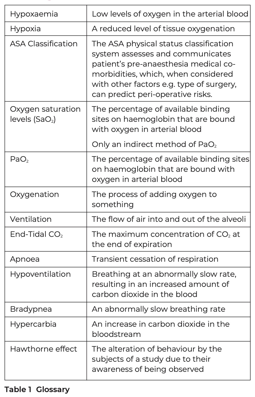
Glossary
Acknowledgements
This essay is based on a submission for my research project submission at Cardiff University, School of Dentistry. The project was supervised by Dr Charlotte Richards, Clinical lecturer/StR Oral Surgery. It is entirely my own work.
References
- Araújo J d O, Bergamaschi C d C, Lopes L C, et al. Effectiveness and safety of oral sedation in adult patients undergoing dental procedures: a systematic review. BMJ Open. 2021;11:e043363. doi:10.1136/bmjopen-2020-043363.
- Scottish Dental Effectiveness Programme (SDCEP). Conscious Sedation In Dentistry: Dental Clinical Guidance 2017 [Available from: Online information available at: https://www.sdcep.org.uk/media/iota3oqm/sdcep-conscious-sedation-guidance-unchanged-2022.pdf
- Kapur A, Kapur V. Conscious Sedation in Dentistry. Ann Maxillofac Surg. 2018;8:320–323. doi:10.4103/ams.ams_191_18.
- Coomar J. Clinical and electronic monitoring during procedural sedation and analgesia for dentistry. SAAD Dig. 2021;37:56-59.
- Weissheimer T, Gerzson A d S, Schwengber H E, Neto A M. Benzodiazepines for conscious sedation in the dental office. Stomatos. 2016;22:42-53.
- Bennett J D, Kramer K J, Bosack R C. How safe is deep sedation or general anesthesia while providing dental care? J Am Dent Assoc. 2015;146:705-708. doi:10.1016/j.adaj.2015.04.005
- Takaya K, Higuchi H, Ishii-Maruhama M, et al. Capnography Prevents Hypoxia during Sedation for Dental Treatment: A Randomized Controlled Trial. JDR Clin Trans Res. 2017;2:158-167. doi:10.1177/2380084416674670.
- Anderson J A, Clark P J, Kafer E R. Use of capnography and transcutaneous oxygen monitoring during outpatient general anesthesia for oral surgery. J Oral Maxillofac Surg. 1987;45:3-10. doi:10.1016/0278-2391(87)90078-4
- Iwasaki J, Vann W F, Jr., Dilley D C, Anderson J A. An investigation of capnography and pulse oximetry as monitors of pediatric patients sedated for dental treatment. Pediatr Dent. 1989;11:111-117.
- Intercollegiate Advisory Committee for Sedation in Dentistry. Standards for Conscious Sedation in the Provision of Dental Care (V1.1). London: Royal Colleges of Surgeons and Royal College of Anaesthetists; 2020.
- Karamnov S, Sarkisian N, Grammer R, Gross W L, Urman R D. Analysis of Adverse Events Associated With Adult Moderate Procedural Sedation Outside the Operating Room. J Patient Saf. 2017;13:111-121. doi:10.1097/pts.0000000000000135.
- Baillard C, Boubaya M, Statescu E, et al. Incidence and risk factors of hypoxaemia after preoxygenation at induction of anaesthesia. Br J Anaesth. 2019;122:388-394. doi:10.1016/j.bja.2018.11.022.
- Academy of Medical Royal Colleges. Safe Sedation Practice for Healthcare Procedures. London; 2013.
- Nagler J M D, Krauss B M D E. Monitoring the Procedural Sedation Patient: Optimal Constructs for Patient Safety. Clin Pediatr Emerg Med. 2010;11:251-264. doi:10.1016/j.cpem.2010.09.007.
- Brady P, Gallagher C, McCarthy C, O'Halloran K, McCreary C, Ioham G. Capnography monitoring during dental conscious sedation Oral Surg. 2016;10:131-136.
- Deitch K, Miner J, Chudnofsky C R, Dominici P, Latta D. Does end tidal CO2 monitoring during emergency department procedural sedation and analgesia with propofol decrease the incidence of hypoxic events? A randomized, controlled trial. Ann Emerg Med. 2010;55:258-264. doi:10.1016/j.annemergmed.2009.07.030.
- Aminiahidashti H, Shafiee S, Zamani Kiasari A, Sazgar M. Applications of End-Tidal Carbon Dioxide (ETCO2) Monitoring in Emergency Department; a Narrative Review. Emerg (Tehran). 2018;6:e5. doi:10.22037/emergency.v6i1.19298.
- Brady P J, Wilson K E, Meeke R, Girdler N M, Mc Creary C. Capnometry monitoring during intravenous sedation with midazolam for oral surgery: Capnometry for conscious sedation in oral surgery. Oral surg. 2015;9:94-101. doi:10.1111/ors.12169.
- Lightdale J R, Goldmann D A, Feldman H A, Newburg A R, DiNardo J A, Fox V L. Microstream Capnography Improves Patient Monitoring During Moderate Sedation: A Randomized, Controlled Trial. Pediatrics. 2006;117:e1170-1178. doi:10.1542/peds.2005-1709.
- Rollert M K, Busaidy K, Krishnan D, Van Heukelom E. Clinical Practice Guidelines for Oral and Maxillofacial Surgery: Anesthesia In Outpatient Facilities. J Oral Maxillofac Surg. 2017;75:e34-49. doi:10.1016/j.joms.2017.04.032.
- American Dental Association House of Delegates. Guidelines for the Use of Sedation and General Anesthesia by Dentists. Illinois; 2016.
- Committee on Standards and Practice Parameters (CSPP). Standards for Basic Anesthetic Monitoring. Illinois; 2020 update.
- Deitch K, Miner J, Chudnofsky C R, Dominici P, Latta D. Does end tidal CO2 monitoring during emergency department procedural sedation and analgesia with propofol decrease the incidence of hypoxic events? A randomized, controlled trial. Ann Emerg Med. 2010;55.
- Deitch K D O, Chudnofsky C R M D, Dominici P M D. The Utility of Supplemental Oxygen During Emergency Department Procedural Sedation With Propofol: A Randomized, Controlled Trial. Ann Emerg Med. 2008;52:1-8. doi:10.1016/j.annemergmed.2007.11.040.
- Klare P, Reiter J, Meining A, et al. Capnographic monitoring of midazolam and propofol sedation during ERCP: a randomized controlled study (EndoBreath Study). Endoscopy. 2016;48:42-50. doi:10.1055/s-0034-1393117.
- Ismail M, Kerr B, Flynn H. Capnography: How much will it cost? A rapid scoping review and heuristic estimate. Paper presented at the Society for the Advancement of Anaethesia in Dentistry symposium, Royal Society of Medicine. Sep 2023.
- Peveling-Oberhag J, Michael F, Tal A, et al. Capnography monitoring of non- anesthesiologist provided sedation during percutaneous endoscopic gastrostomy placement: A prospective, controlled, randomized trial. J Gastroenterol Hepatol. 2020;35:401-407. doi:10.1111/jgh.14760 .
- Ishiwata T, Tsushima K, Terada J, et al. Efficacy of End-Tidal Capnography Monitoring during Flexible Bronchoscopy in Nonintubated Patients under Sedation: A Randomized Controlled Study. Respiration. 2018;96:355-362. doi:10.1159/000489888.
- Brady P, Iohom G, O'Halloran K D, McCreary C, Cronin M. Microstream capnography during conscious sedation with midazolam for oral surgery: a randomised controlled trial. BDJ Open. 2017;3:17019. doi:10.1038/bdjopen.2017.19.
- Mehta P P, Kochhar G, Albeldawi M, et al. Capnographic Monitoring in Routine EGD and Colonoscopy With Moderate Sedation: A Prospective, Randomized, Controlled Trial. Am J Gastroenterol. 2016;111:395-404. doi:10.1038/ajg.2015.437.
- Langhan M L, Shabanova V, Li F Y, Bernstein S L, Shapiro E D. A randomized controlled trial of capnography during sedation in a pediatric emergency setting. Am J Emerg Med. 2015;33:25-30. doi:10.1016/j.ajem.2014.09.050 .
- Zongming J, Zhonghua C, Xiangming F. Sidestream capnographic monitoring reduces the incidence of arterial oxygen desaturation during propofol ambulatory anesthesia for surgical abortion. Med Sci Monit. 2014;20:2336-2342. doi:10.12659/msm.891023.
- Beitz A, Riphaus A, Meining A, et al. Capnographic monitoring reduces the incidence of arterial oxygen desaturation and hypoxemia during propofol sedation for colonoscopy: a randomized, controlled study (ColoCap Study). Am J Gastroenterol. 2012;107:1205-1212. doi:10.1038/ajg.2012.136.
- Qadeer M A, Vargo J J, Dumot J A, et al. Capnographic monitoring of respiratory activity improves safety of sedation for endoscopic cholangiopancreatography and ultrasonography. Gastroenterology. 2009;136:1568-1576. doi:10.1053/j.gastro.2009.02.004.
- Vargo J J, Zuccaro G, Dumot J A, Conwell D L, Morrow J B, Shay S S. Automated graphic assessment of respiratory activity is superior to pulse oximetry and visual assessment for the detection of early respiratory depression during therapeutic upper endoscopy. Gastrointest Endosc. 2002;55:826-831. doi:10.1067/mge.2002.124208.
- Lightdale J R, Goldmann D A, Feldman H A, Newburg A R, DiNardo J A, Fox V L. Microstream Capnography Improves Patient Monitoring During Moderate Sedation: A Randomized, Controlled Trial. Pediatrics (Evanston). 2006;117:e1170- e1178. doi:10.1542/peds.2005-1709.
- Fu E S, Downs J B, Schweiger J W, Miguel R V, Smith R A. Supplemental oxygen impairs detection of hypoventilation by pulse oximetry. Chest. 2004;126:1552- 1558. doi: 10.1378/chest.126.5.1552.
- Ishiwata T, Tsushima K, Fujie M, et al. End-tidal capnographic monitoring to detect apnea episodes during flexible bronchoscopy under sedation. BMC Pulm Med. 2017;17:7. doi:10.1186/s12890-016-0361-7.
- 39. Sivilotti M L A, Messenger D W, Van Vlymen J, Dungey P E, Murray H E. A comparative evaluation of capnometry versus pulse oximetry during procedural sedation and analgesia on room air. CJEM. 2010;12:397-404. doi:10.1017/S1481803500012549.
- 40. Waugh J B, Epps C A, Khodneva Y A. Capnography enhances surveillance of respiratory events during procedural sedation: a meta-analysis. J Clin Anesth. 2011;23:189-196. doi:10.1016/j.jclinane.2010.08.012 .
- 41. Long B, Koyfman A, Vivirito M A. Capnography in the Emergency Department: A Review of Uses, Waveforms, and Limitations. J Emerg Med. 2017;53:829-842. doi:10.1016/j.jemermed.2017.08.026.